中国全科医学 ›› 2022, Vol. 25 ›› Issue (09): 1077-1081.DOI: 10.12114/j.issn.1007-9572.2021.02.026

所属专题: 指南/共识最新文章合集

• 论著 • 上一篇    下一篇

甲状腺结节超声恶性风险分层方法对甲状腺结节良恶性的鉴别诊断价值:中美指南对比分析

李健*, 殷延华, 戚建国, 王洲, 任永凤, 王珊珊, 张伟莉, 王效军, 唐坤   

  1. 236800 安徽省亳州市人民医院超声科
  • 收稿日期:2021-06-09 修回日期:2021-10-17 出版日期:2022-03-20 发布日期:2022-03-01
  • 通讯作者: 李健

Ultrasound-based Thyroid Nodule Malignancy Risk Stratification in Differentiating Benign and Malignant Thyroid Nodulesa Comparative Analysis between the C-TIRADS and ATA Guidelines

LI Jian*YIN YanhuaQI JianguoWANG ZhouREN YongfengWANG ShanshanZHANG WeiliWANG XiaojunTANG Kun   

  1. LI Jian*YIN YanhuaQI JianguoWANG ZhouREN YongfengWANG ShanshanZHANG WeiliWANG XiaojunTANG Kun

  • Received:2021-06-09 Revised:2021-10-17 Published:2022-03-20 Online:2022-03-01

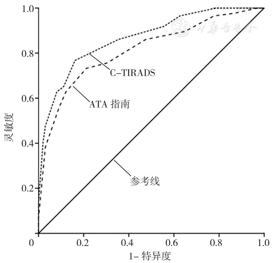
摘要: 背景2020年发布的《甲状腺结节超声恶性危险分层中国指南》(简称C-TIRADS)提出了甲状腺结节超声恶性风险分层方法,但目前关于其对甲状腺结节良恶性鉴别诊断价值的研究较少。目的探讨C-TIRADS与2015年美国甲状腺学会(ATA)发布的《成人甲状腺结节与分化型甲状腺癌诊治指南》(简称ATA指南)中甲状腺结节超声恶性风险分层方法对甲状腺结节良恶性的鉴别诊断价值。方法选取2019年1月至2021年5月在亳州市人民医院住院并行手术治疗和超声检查的甲状腺结节患者336例为研究对象。选取4名具有主治医师以上职称的超声科医师并随机分为A组和B组,每组2名;A组、B组超声科医师分别依据C-TIRADS、ATA指南中甲状腺结节超声恶性风险分层方法对所有患者超声检查结果进行评估。以术中或术后病理检查结果为"金标准",绘制受试者工作特征(ROC)曲线以分析C-TIRADS、ATA指南中甲状腺结节超声恶性风险分层方法对甲状腺结节良恶性的鉴别诊断价值。结果本组336例甲状腺结节患者经术中或术后病理检查共发现367个甲状腺结节,其中良性结节253个、恶性结节114个。A组超声科医师将良性结节评估为5类2个、4c类23个、4b类30个、4a类80个、3类103个、2类15个,将恶性结节评估为5类24个、4c类59个、4b类22个、4a类6个、3类3个、2类0个;B组超声科医师将良性结节评估为高度可疑恶性13个、中度可疑恶性53个、低度可疑恶性47个、极低度可疑恶性118个、良性结节22个,将恶性结节评估为高度可疑恶性76个、中度可疑恶性24个、低度可疑恶性8个、极低度可疑恶性6个、良性结节0个。C-TIRADS、ATA指南中甲状腺结节超声恶性风险分层方法鉴别诊断甲状腺结节良恶性的ROC曲线下面积分别为0.890〔95%CI(0.815,0.918)〕、0.780〔95%CI(0.750,0.876)〕,差异有统计学意义(Z=13.62,P<0.05)。依据C-TIRADS中甲状腺结节超声恶性风险分层方法,4b类、4a类鉴别诊断甲状腺结节良恶性的灵敏度分别为92.11%、97.37%,特异度分别为78.26%、46.64%,准确度分别为82.56%、62.40%,约登指数分别为0.70、0.44;依据ATA指南中甲状腺结节超声恶性风险分层方法,中度可疑恶性、低度可疑恶性鉴别诊断甲状腺结节良恶性的灵敏度分别为87.72%、94.74%,特异度分别为67.98%、44.66%,准确度分别为74.11%、60.22%,约登指数分别为0.56、0.39。结论C-TIRADS中甲状腺结节超声恶性风险分层方法对甲状腺结节良恶性的鉴别诊断价值高于ATA指南中的方法,且最佳截断值为4b类。

关键词: 甲状腺结节, 诊断, 鉴别, 超声检查, 指南, 对比研究

Abstract: Background

The 2020 Chinese guidelines for ultrasound malignancy risk stratification of thyroid nodules: the C-TIRADS (C-TIRADS for short) put forward a method for stratifying the malignancy risk of thyroid nodules, but there are few studies about its value in differentiating benign and malignant thyroid nodules.

Objective

To compare the value of ultrasound-based thyroid nodule malignancy risk stratification in differentiating malignant and benign thyroid nodule between C-TIRADS and 2015 American Thyroid Association Management Guidelines for Adult Patients with Thyroid Nodules and Differentiated Thyroid Cancer: the American Thyroid Association Guidelines Task Force on Thyroid Nodules and Differentiated Thyroid Cancer (ATA guidelines for short) .

Methods

A total of 336 inpatients who underwent surgery and ultrasound examination in Bozhou People's Hospital from January 2019 to May 2021 were selected as participants. Four ultrasonologists with a title higher than attending physician were equally randomized into groups A and B for evaluating ultrasonic examination results according to ultrasound-based thyroid nodule malignancy risk stratification in C-TIRADS and ATA guidelines, respectively. The accuracies of ultrasound-based thyroid nodule malignancy stratification in the two guidelines in differentiating malignant and benign thyroid nodules were assessed using intraoperative or postoperative pathological findings (gold standard for diagnosis) , and were further analyzed using ROC analysis.

Results

According to intraoperative or postoperative pathological findings, the participants had 367 thyroid nodules in total, including 253 benign nodules and 114 malignant nodules. The nodules were classified as follows according to the TI-RADS in the C-TIRADS by ultrasonologists in group A: (1) 253 benign nodules: 2 were in TI-RADS 5 category, 23, 30, and 80 were in categories 4c, 4b, and 4a, respectively, 103 were in 3 category, and 15 were in 2 category; (2) 114 malignant nodules: 24 were in TI-RADS 5 category, 59 were in 4c category, 22 were in 4b category, 6 were in 4a category and 3 were in 3 category. And the classification of nodules according to the ATA guideline by ultrasonologists in group B was: (1) 253 benign nodules: 13, 53, 47, 118 were high, intermediate, low, and very low suspicion pattern for malignancy, respectively, and 22 were benign pattern; (2) 114 malignant nodules: 76, 24, 8, 6 were high, intermediate, low, and very low suspicion pattern for malignancy, respectively. ROC analysis showed that, AUCs of ultrasound-based thyroid nodule malignancy risk stratification in differentiating malignant and benign thyroid nodules between C-TIRADS and ATA guidelines were 0.890〔95%CI (0.815, 0.918) 〕and 0.780〔95%CI (0.750, 0.876) 〕, with significant difference (Z=13.62, P<0.05) . The TI-RADS in the C-TIRADS showed 92.11% sensitivity, 78.26% specificity, 82.56% accuracy, with Youden index of 0.70 in differentiating 4b category benign and malignant nodules, and demonstrated 97.37% sensitivity, 46.64% specificity, 62.40% accuracy with Youden index of 0.44 in differentiating 4a category benign and malignant nodules. The ultrasound-based thyroid nodule malignancy risk stratification in ATA guideline showed 87.72% sensitivity, 67.98% specificity, 74.11% accuracy, with Youden index of 0.56 in differentiating intermediate suspicion pattern for malignancy, and demonstrated 94.74% sensitivity, 44.66% specificity, 60.22% accuracy with Youden index of 0.39 in differentiating low suspicion pattern for malignancy.

Conclusion

The ultrasound-based thyroid nodule malignancy risk stratification in C-TIRADS has higher value in differentiating malignant and benign thyroid nodules than that in ATA guidelines, and the optimal performance of which may be manifested in differentiating malignant and benign thyroid nodules of TI-RADS 4b category.

Key words: Thyroid nodule, Diagnosis, differential, Ultrasonography, Guidebook, Comparative study

中图分类号: