
中国全科医学 ›› 2026, Vol. 29 ›› Issue (35): 5290-5299.DOI: 10.12114/j.issn.1007-9572.2025.0398
李俊杰1,2, 梁俊荣1, 刘震雄1, 王新1, 窦维佳1,*( )
收稿日期:2025-09-30
修回日期:2025-11-25
出版日期:2026-12-15
发布日期:2026-10-26
通讯作者:
窦维佳
作者贡献:
李俊杰进行文章的构思与设计,Meta分析的结果解读,撰写文章;李俊杰、梁俊荣负责检索策略的制定、文献筛选、风险偏倚评估;窦维佳负责文章的质量控制及审校;刘震雄、王新、窦维佳负责项目的牵头和文章的修订;李俊杰、窦维佳对文章整体负责。
基金资助:
LI Junjie1,2, LIANG Junrong1, LIU Zhenxiong1, WANG Xin1, DOU Weijia1,*()
Received:2025-09-30
Revised:2025-11-25
Published:2026-12-15
Online:2026-10-26
Contact:
DOU Weijia
摘要: 背景 浅表食管癌(SEC)浸润深度诊断与治疗预后息息相关,目前尚无关于SEC浸润深度诊断的最佳方法共识。 目的 系统评价光学相干断层扫描(OCT)、放大内镜(ME)以及超声内镜(EUS)诊断SEC黏膜上皮层/黏膜固有层(EP/LPM)病变的诊断效能。 方法 计算机检索PubMed、Embase、Web of Science、Cochrane Library等英文数据库以及中国知网(CNKI)、中国生物医学文献服务系统(SinoMed)、维普网(VIP)、万方数据知识服务平台(Wanfang Data)等中文数据库中关于OCT、ME以及EUS诊断SEC EP/LPM病变的研究文献,检索时限从建库至2024年10月。由2名研究者独立筛选文献、提取资料并评价纳入研究的偏倚风险,采用Stata 16.0、RevMan 5.3、Meta-Disc 1.4及R 4.4.0等软件进行Meta分析。 结果 共纳入21项诊断性试验,Meta分析结果显示,OCT、EUS和ME诊断SEC EP/LPM病变的灵敏度、特异度和准确度分别为OCT:0.93(95%CI=0.89~0.96)、0.77(95%CI=0.54~0.91)、0.87(95%CI=0.74~0.94),EUS:0.73(95%CI=0.62~0.82)、0.82(95%CI=0.72~0.89)、0.77(95%CI=0.72~0.81),ME:0.86(95%CI=0.81~0.90)、0.75(95%CI=0.62~0.84)、0.82(95%CI=0.78~0.85);OCT、EUS和ME诊断SEC EP/LPM病变的受试者工作特征曲线下面积分别为0.92(95%CI=0.86~0.98)、0.83(95%CI=0.80~0.86)和0.86(95%CI=0.85~0.88)。Deeks漏斗图结果显示,OCT、EUS、ME研究散点对称性分布于回归线的两侧,说明无发表偏倚(P均>0.05)。 结论 OCT和ME在诊断SEC EP/LPM病变方面展现出较高的灵敏度与准确度,其综合诊断效能优于EUS。
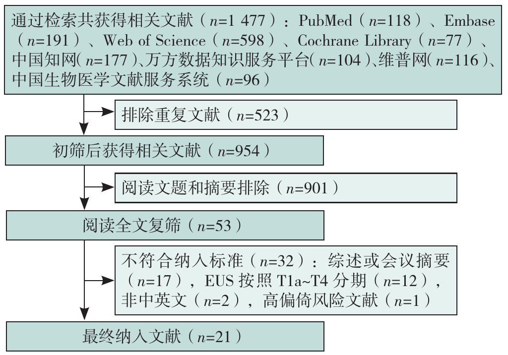
图1 文献筛选流程图注:EUS=超声内镜。
Figure 1 Literature screening flowchart
| 第一作者 | 发表年份(年) | 国家 | 研究类型 | 参与单位 | 年龄( | 成像方式 | 放大分型 | 探头种类及频率(MHz) | 病变数量(个) | TP | FP | FN | TN |
|---|---|---|---|---|---|---|---|---|---|---|---|---|---|
| Mizumoto[ | 2018 | 日本 | R | S | 67.7±8.8 | EUS | — | 径向探头20 | 174 | 89 | 17 | 35 | 33 |
| ME-NBI | JES | — | 174 | 103 | 11 | 21 | 39 | ||||||
| Mizumoto[ | 2018 | 日本 | R | S | 68.2±8.9 | ME-NBI | JES | — | 136 | 72 | 11 | 15 | 38 |
| Esaki[ | 2006 | 日本 | P | S | 66.0±7.8 | EUS | — | 微探头20 | 40 | 14 | 1 | 7 | 18 |
| Hatta[ | 2010 | 日本 | P | S | 66.3±10.5 | OCT | — | — | 109 | 74 | 3 | 4 | 28 |
| Hatta[ | 2012 | 日本 | P | S | 68.0±8.2 | OCT | — | — | 131 | 88 | 8 | 5 | 30 |
| EUS | — | 微探头20 | 131 | 75 | 12 | 18 | 26 | ||||||
| Zeng[ | 2022 | 中国 | R | S | 61.3±7.5 | EUS | — | 径向/微探头7.5~20 | 152 | 57 | 14 | 21 | 60 |
| ME-NBI | JES | — | 152 | 66 | 6 | 12 | 68 | ||||||
| 向京元[ | 2019 | 中国 | P | S | 60.5±7.7 | OCT | — | — | 28 | 15 | 5 | 4 | 4 |
| ME-NBI | JES | — | 28 | 13 | 1 | 6 | 8 | ||||||
| Ebi[ | 2015 | 日本、美国 | P | M | 68.0±5.9 | ME-BLI | Inoue | — | 49 | 21 | 7 | 10 | 11 |
| Kim[ | 2017 | 韩国 | R | M | 66.0±6.8 | ME-NBI | JES | — | 70 | 20 | 0 | 8 | 42 |
| Oyama[ | 2017 | 日本 | P | M | — | ME-BLI | JES | — | 211 | 159 | 13 | 4 | 35 |
| Fujiyoshi[ | 2017 | 日本 | P | S | 67.0±9.6 | ME-NBI | JES | — | 145 | 92 | 21 | 10 | 22 |
| ME-NBI | Inoue | — | 145 | 94 | 28 | 8 | 15 | ||||||
| ME-NBI | Arima | — | 145 | 102 | 31 | 0 | 12 | ||||||
| Diao[ | 2018 | 中国 | P | S | 62.0±7.2 | ME-BLI | JES | — | 42 | 12 | 1 | 2 | 27 |
| Katada[ | 2019 | 日本 | R | S | 71.0±9.0 | ME-NBI | JES | — | 256 | 156 | 11 | 16 | 73 |
| Hatta[ | 2021 | 日本 | P | S | 68.4±9.4 | ME-NBI | JES | — | 81 | 55 | 9 | 9 | 8 |
| ME-BLI | JES | — | 81 | 54 | 10 | 10 | 7 | ||||||
| Suzuki[ | 2023 | 日本 | R | S | 69.0±9.6 | ME-NBI | JES | — | 403 | 107 | 39 | 45 | 212 |
| EUS | — | 微探头20 | 403 | 103 | 260 | 49 | 225 | ||||||
| Zhang[ | 2024 | 中国 | R | S | 66.3±7.1 | ME-NBI | JES | — | 338 | 234 | 40 | 35 | 29 |
| 郑永胜[ | 2019 | 中国 | R | S | 67.0±6.2 | EUS | — | 微探头 | 105 | 67 | 11 | 6 | 21 |
| ME-NBI | JES | — | 105 | 69 | 10 | 4 | 22 | ||||||
| 李雪[ | 2022 | 中国 | R | S | 66.2±7.3 | ME-NBI | JES | — | 203 | 94 | 25 | 49 | 35 |
| 袁小志[ | 2022 | 中国 | R | S | 41.7±2.4 | EUS | — | 环扫10 | 109 | 24 | 3 | 35 | 47 |
| ME-NBI | Inoue | — | 109 | 48 | 4 | 11 | 46 | ||||||
| 刘黎[ | 2024 | 中国 | R | S | 55.5±10.6 | ME-BLI | JES | — | 114 | 64 | 5 | 14 | 31 |
| 汪波[ | 2022 | 中国 | R | S | — | EUS | — | 微探头20 | 54 | 36 | 1 | 11 | 6 |
表1 纳入研究的基本特征
Table 1 Basic characteristics of included studies
| 第一作者 | 发表年份(年) | 国家 | 研究类型 | 参与单位 | 年龄( | 成像方式 | 放大分型 | 探头种类及频率(MHz) | 病变数量(个) | TP | FP | FN | TN |
|---|---|---|---|---|---|---|---|---|---|---|---|---|---|
| Mizumoto[ | 2018 | 日本 | R | S | 67.7±8.8 | EUS | — | 径向探头20 | 174 | 89 | 17 | 35 | 33 |
| ME-NBI | JES | — | 174 | 103 | 11 | 21 | 39 | ||||||
| Mizumoto[ | 2018 | 日本 | R | S | 68.2±8.9 | ME-NBI | JES | — | 136 | 72 | 11 | 15 | 38 |
| Esaki[ | 2006 | 日本 | P | S | 66.0±7.8 | EUS | — | 微探头20 | 40 | 14 | 1 | 7 | 18 |
| Hatta[ | 2010 | 日本 | P | S | 66.3±10.5 | OCT | — | — | 109 | 74 | 3 | 4 | 28 |
| Hatta[ | 2012 | 日本 | P | S | 68.0±8.2 | OCT | — | — | 131 | 88 | 8 | 5 | 30 |
| EUS | — | 微探头20 | 131 | 75 | 12 | 18 | 26 | ||||||
| Zeng[ | 2022 | 中国 | R | S | 61.3±7.5 | EUS | — | 径向/微探头7.5~20 | 152 | 57 | 14 | 21 | 60 |
| ME-NBI | JES | — | 152 | 66 | 6 | 12 | 68 | ||||||
| 向京元[ | 2019 | 中国 | P | S | 60.5±7.7 | OCT | — | — | 28 | 15 | 5 | 4 | 4 |
| ME-NBI | JES | — | 28 | 13 | 1 | 6 | 8 | ||||||
| Ebi[ | 2015 | 日本、美国 | P | M | 68.0±5.9 | ME-BLI | Inoue | — | 49 | 21 | 7 | 10 | 11 |
| Kim[ | 2017 | 韩国 | R | M | 66.0±6.8 | ME-NBI | JES | — | 70 | 20 | 0 | 8 | 42 |
| Oyama[ | 2017 | 日本 | P | M | — | ME-BLI | JES | — | 211 | 159 | 13 | 4 | 35 |
| Fujiyoshi[ | 2017 | 日本 | P | S | 67.0±9.6 | ME-NBI | JES | — | 145 | 92 | 21 | 10 | 22 |
| ME-NBI | Inoue | — | 145 | 94 | 28 | 8 | 15 | ||||||
| ME-NBI | Arima | — | 145 | 102 | 31 | 0 | 12 | ||||||
| Diao[ | 2018 | 中国 | P | S | 62.0±7.2 | ME-BLI | JES | — | 42 | 12 | 1 | 2 | 27 |
| Katada[ | 2019 | 日本 | R | S | 71.0±9.0 | ME-NBI | JES | — | 256 | 156 | 11 | 16 | 73 |
| Hatta[ | 2021 | 日本 | P | S | 68.4±9.4 | ME-NBI | JES | — | 81 | 55 | 9 | 9 | 8 |
| ME-BLI | JES | — | 81 | 54 | 10 | 10 | 7 | ||||||
| Suzuki[ | 2023 | 日本 | R | S | 69.0±9.6 | ME-NBI | JES | — | 403 | 107 | 39 | 45 | 212 |
| EUS | — | 微探头20 | 403 | 103 | 260 | 49 | 225 | ||||||
| Zhang[ | 2024 | 中国 | R | S | 66.3±7.1 | ME-NBI | JES | — | 338 | 234 | 40 | 35 | 29 |
| 郑永胜[ | 2019 | 中国 | R | S | 67.0±6.2 | EUS | — | 微探头 | 105 | 67 | 11 | 6 | 21 |
| ME-NBI | JES | — | 105 | 69 | 10 | 4 | 22 | ||||||
| 李雪[ | 2022 | 中国 | R | S | 66.2±7.3 | ME-NBI | JES | — | 203 | 94 | 25 | 49 | 35 |
| 袁小志[ | 2022 | 中国 | R | S | 41.7±2.4 | EUS | — | 环扫10 | 109 | 24 | 3 | 35 | 47 |
| ME-NBI | Inoue | — | 109 | 48 | 4 | 11 | 46 | ||||||
| 刘黎[ | 2024 | 中国 | R | S | 55.5±10.6 | ME-BLI | JES | — | 114 | 64 | 5 | 14 | 31 |
| 汪波[ | 2022 | 中国 | R | S | — | EUS | — | 微探头20 | 54 | 36 | 1 | 11 | 6 |
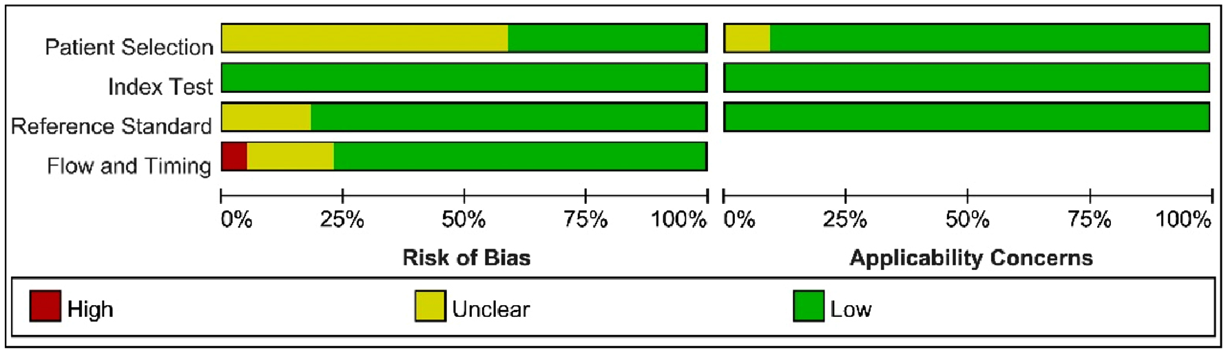
图2 纳入研究偏倚风险总结图
Figure 2 Summary of bias risk for included studies
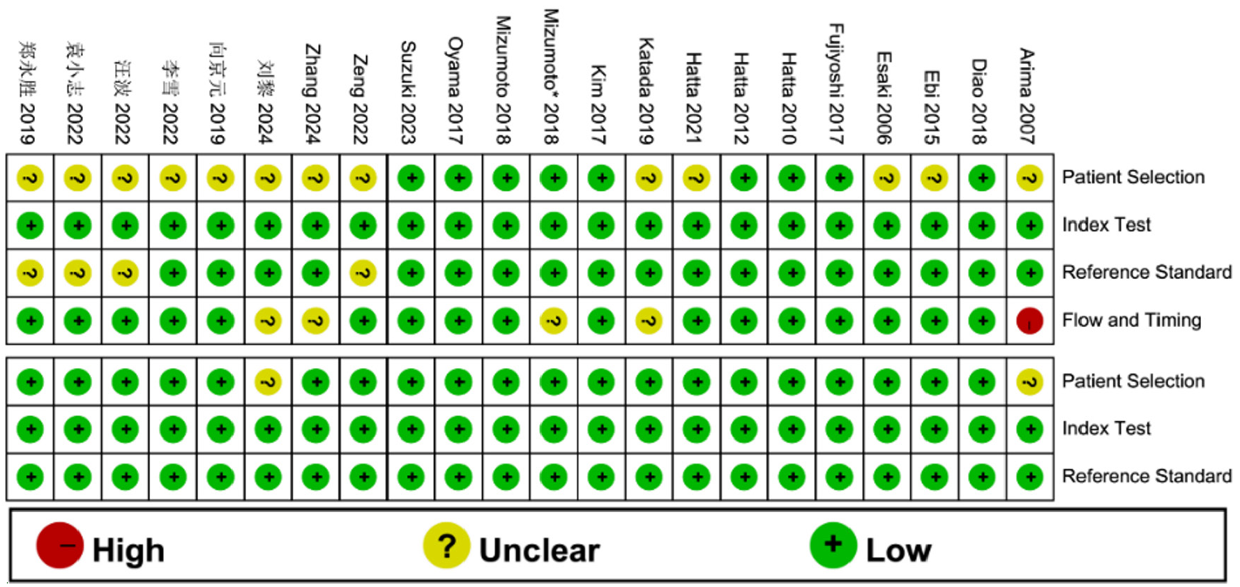
图3 纳入研究偏倚风险评估图
Figure 3 Risk of bias assessment graph for included studies
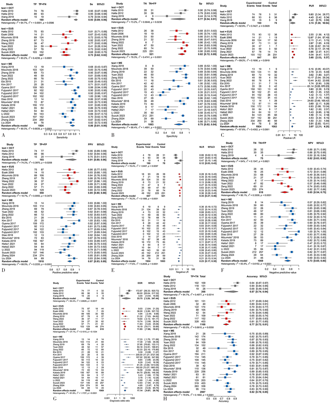
图4 不同成像方式诊断SEC EP/LPM病变的森林图注:A为灵敏度,B为特异度,C为阳性似然比,D为阳性预测值,E为阴性似然比,F为阴性预测值,G为诊断比值比,H为准确度;TP=真阳性,FP=假阳性,FN=假阴性,TN=真阴性,OCT=光学相干断层扫描,ME=放大内镜,PPV=阳性预测值,NPV=阴性预测值,PLR=阳性似然比,NLR=阴性似然比,DOR=诊断比值比。
Figure 4 Summary forest plot of diagnostic accuracy for SEC EP/LPM lesions using different imaging modalities
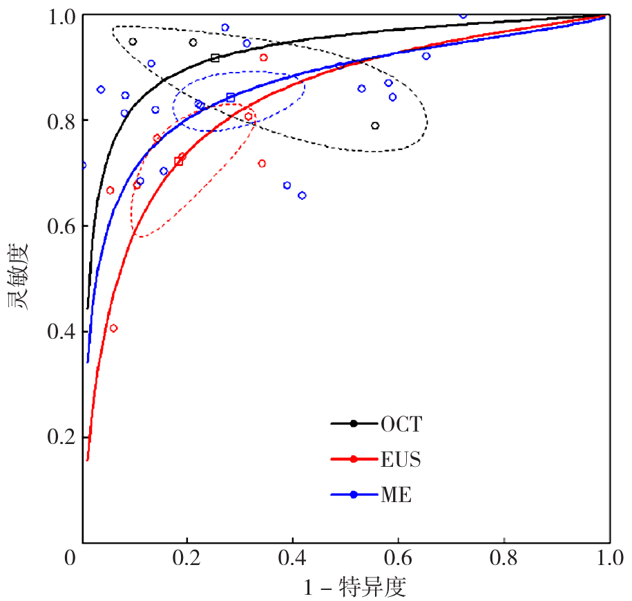
图5 不同成像方式诊断SEC EP/LPM病变的SROC曲线
Figure 5 SROC curves for diagnosing SEC EP/LPM lesions using different imaging modalities
| 协变量 | 文献数量(篇) | 灵敏度(95%CI) | Z值 | P值 | 特异度(95%CI) | Z值 | P值 |
|---|---|---|---|---|---|---|---|
| 探头类型 | 3.21 | <0.01 | 1.22 | 0.22 | |||
| 环扫 | 1[ | 0.41(0.21~0.60) | 0.94(0.86~1.00) | ||||
| 微探头/径向 | 7[ | 0.76(0.70~0.82) | 0.79(0.71~0.87) | ||||
| 研究类型 | 0.15 | 0.58 | 0.62 | 0.53 | |||
| 前瞻性研究 | 2[ | 0.74(0.55~0.93) | 0.82(0.63~1.00) | ||||
| 回顾性研究 | 6[ | 0.72(0.61~0.83) | 0.83(0.73~0.92) | ||||
| 发表年份 | 0.19 | 0.85 | 3.29 | <0.01 | |||
| 2020年前 | 4[ | 0.78(0.68~0.89) | 0.71(0.61~0.82) | ||||
| 2020年及以后 | 4[ | 0.66(0.52~0.79) | 0.89(0.84~0.94) | ||||
| 研究国家 | 1.08 | 0.28 | 0.31 | 0.25 | |||
| 日本 | 4[ | 0.72(0.58~0.86) | 0.82(0.69~0.94) | ||||
| 非日本 | 4[ | 0.73(0.59~0.87) | 0.84(0.71~0.96) | ||||
| 病变数量 | 1.00 | 0.50 | 0.97 | 0.33 | |||
| <100个 | 2[ | 0.73(0.52~0.93) | 0.92(0.79-1.00) | ||||
| ≥100个 | 6[ | 0.73(0.62~0.84) | 0.81(0.71~0.91) |
表2 EUS研究的Meta回归分析结果
Table 2 Meta-regression analysis results for EUS studies
| 协变量 | 文献数量(篇) | 灵敏度(95%CI) | Z值 | P值 | 特异度(95%CI) | Z值 | P值 |
|---|---|---|---|---|---|---|---|
| 探头类型 | 3.21 | <0.01 | 1.22 | 0.22 | |||
| 环扫 | 1[ | 0.41(0.21~0.60) | 0.94(0.86~1.00) | ||||
| 微探头/径向 | 7[ | 0.76(0.70~0.82) | 0.79(0.71~0.87) | ||||
| 研究类型 | 0.15 | 0.58 | 0.62 | 0.53 | |||
| 前瞻性研究 | 2[ | 0.74(0.55~0.93) | 0.82(0.63~1.00) | ||||
| 回顾性研究 | 6[ | 0.72(0.61~0.83) | 0.83(0.73~0.92) | ||||
| 发表年份 | 0.19 | 0.85 | 3.29 | <0.01 | |||
| 2020年前 | 4[ | 0.78(0.68~0.89) | 0.71(0.61~0.82) | ||||
| 2020年及以后 | 4[ | 0.66(0.52~0.79) | 0.89(0.84~0.94) | ||||
| 研究国家 | 1.08 | 0.28 | 0.31 | 0.25 | |||
| 日本 | 4[ | 0.72(0.58~0.86) | 0.82(0.69~0.94) | ||||
| 非日本 | 4[ | 0.73(0.59~0.87) | 0.84(0.71~0.96) | ||||
| 病变数量 | 1.00 | 0.50 | 0.97 | 0.33 | |||
| <100个 | 2[ | 0.73(0.52~0.93) | 0.92(0.79-1.00) | ||||
| ≥100个 | 6[ | 0.73(0.62~0.84) | 0.81(0.71~0.91) |
| 协变量 | 文献数量(篇) | 灵敏度(95%CI) | Z值 | P值 | 特异度(95%CI) | Z值 | P值 |
|---|---|---|---|---|---|---|---|
| 增强方式 | 1.88 | 0.06 | 0.34 | 0.73 | |||
| BLI | 5[ | 0.87(0.77~0.96) | 0.77(0.57~0.97) | ||||
| NBI | 12[ | 0.86(0.80~0.91) | 0.74(0.62~0.87) | ||||
| 放大分型 | 2.58 | <0.01 | 0.81 | 0.42 | |||
| JES | 14[ | 0.85(0.79~0.90) | 0.78(0.68~0.88) | ||||
| Arima/Inoue | 3[ | 0.90(0.83~0.98) | 0.58(0.29~0.87) | ||||
| 参与单位 | 1.26 | 0.21 | 1.28 | 0.20 | |||
| 单中心 | 14[ | 0.86(0.81~0.91) | 0.72(0.60~0.84) | ||||
| 多中心 | 3[ | 0.86(0.73~0.99) | 0.86(0.69~1.00) | ||||
| 研究类型 | 1.88 | 0.06 | 2.58 | <0.01 | |||
| 前瞻性研究 | 6[ | 0.90(0.85~0.95) | 0.60(0.43~0.78) | ||||
| 回顾性研究 | 11[ | 0.83(0.76~0.90) | 0.83(0.73~0.92) | ||||
| 发表年份 | 1.75 | 0.08 | 0.52 | 0.62 | |||
| 2020年前 | 10[ | 0.89(0.84~0.94) | 0.76(0.63~0.90) | ||||
| 2020年及以后 | 7[ | 0.81(0.72~0.89) | 0.73(0.55~0.90) | ||||
| 研究国家 | 1.75 | 0.08 | 2.58 | <0.01 | |||
| 日本 | 8[ | 0.89(0.84~0.94) | 0.63(0.48~0.79) | ||||
| 非日本 | 9[ | 0.82(0.73~0.90) | 0.86(0.76~0.96) | ||||
| 病变数量 | 3.29 | <0.01 | 0.05 | 0.96 | |||
| <100个 | 5[ | 0.78(0.66~0.90) | 0.81(0.65~0.98) | ||||
| ≥100个 | 12[ | 0.88(0.84~0.93) | 0.72(0.59~0.85) |
表3 ME研究的Meta回归分析结果
Table 3 Meta-regression analysis results for ME studies
| 协变量 | 文献数量(篇) | 灵敏度(95%CI) | Z值 | P值 | 特异度(95%CI) | Z值 | P值 |
|---|---|---|---|---|---|---|---|
| 增强方式 | 1.88 | 0.06 | 0.34 | 0.73 | |||
| BLI | 5[ | 0.87(0.77~0.96) | 0.77(0.57~0.97) | ||||
| NBI | 12[ | 0.86(0.80~0.91) | 0.74(0.62~0.87) | ||||
| 放大分型 | 2.58 | <0.01 | 0.81 | 0.42 | |||
| JES | 14[ | 0.85(0.79~0.90) | 0.78(0.68~0.88) | ||||
| Arima/Inoue | 3[ | 0.90(0.83~0.98) | 0.58(0.29~0.87) | ||||
| 参与单位 | 1.26 | 0.21 | 1.28 | 0.20 | |||
| 单中心 | 14[ | 0.86(0.81~0.91) | 0.72(0.60~0.84) | ||||
| 多中心 | 3[ | 0.86(0.73~0.99) | 0.86(0.69~1.00) | ||||
| 研究类型 | 1.88 | 0.06 | 2.58 | <0.01 | |||
| 前瞻性研究 | 6[ | 0.90(0.85~0.95) | 0.60(0.43~0.78) | ||||
| 回顾性研究 | 11[ | 0.83(0.76~0.90) | 0.83(0.73~0.92) | ||||
| 发表年份 | 1.75 | 0.08 | 0.52 | 0.62 | |||
| 2020年前 | 10[ | 0.89(0.84~0.94) | 0.76(0.63~0.90) | ||||
| 2020年及以后 | 7[ | 0.81(0.72~0.89) | 0.73(0.55~0.90) | ||||
| 研究国家 | 1.75 | 0.08 | 2.58 | <0.01 | |||
| 日本 | 8[ | 0.89(0.84~0.94) | 0.63(0.48~0.79) | ||||
| 非日本 | 9[ | 0.82(0.73~0.90) | 0.86(0.76~0.96) | ||||
| 病变数量 | 3.29 | <0.01 | 0.05 | 0.96 | |||
| <100个 | 5[ | 0.78(0.66~0.90) | 0.81(0.65~0.98) | ||||
| ≥100个 | 12[ | 0.88(0.84~0.93) | 0.72(0.59~0.85) |
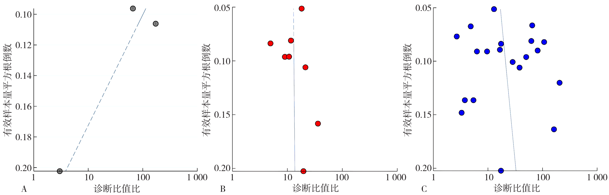
图6 发表偏倚检测Deeks漏斗图注:A为OCT,B为EUS,C为ME。
Figure 6 Deeks funnel plot for publication bias detection
| [1] |
|
| [2] |
|
| [3] |
|
| [4] |
|
| [5] |
|
| [6] |
|
| [7] |
|
| [8] |
|
| [9] |
向京元,令狐恩强,李隆松,等. 光学相干断层扫描技术判断早期食管癌浸润深度的前瞻性研究[J]. 中华消化内镜杂志, 2019, 36(11): 802-805. DOI: 10.3760/cma.j.issn.1007-5232.2019.11.002.
|
| [10] |
|
| [11] |
|
| [12] |
|
| [13] |
|
| [14] |
|
| [15] |
|
| [16] |
|
| [17] |
|
| [18] |
|
| [19] |
郑永胜,雷天霞,杨炜琳,等. 放大内镜窄带光成像联合内镜超声对早期食管癌浸润深度诊断的准确性分析[J]. 中华消化内镜杂志, 2019, 36(10): 761-763.
|
| [20] |
李雪,邢洁,张倩,等. 窄带成像技术联合放大内镜JES分型对早期食管癌的诊断价值[J]. 首都医科大学学报, 2022, 43(2): 210-215. DOI: 10.3969/j.issn.1006-7795.2022.02.008.
|
| [21] |
袁小志,杨瑞娜,董彩红,等. 超声内镜对早期食管癌局部浸润深度的诊断价值[J]. 食管疾病, 2022, 4(3): 214-217. DOI: 10.15926/j.cnki.issn2096-7381.2022.03.011.
|
| [22] |
刘黎,魏文俊. BLI-ME对早期食管癌的诊断价值及B2型血管对浸润深度的判断[J]. 蚌埠医学院学报, 2024, 49(5): 619-622. DOI: 10.13898/j.cnki.issn.1000-2200.2024.05.014.
|
| [23] |
汪波. 放大内镜与超声内镜在诊断早期食管癌浸润深度时的价值探讨[J]. 中文科技期刊数据库(全文版)医药卫生, 2022(2): 194-196.
|
| [24] |
|
| [25] |
|
| [26] |
|
| [27] |
|
| [28] |
|
| [29] |
|
| [30] |
|
| [31] |
|
| [32] |
|
| [33] |
|
| [34] |
|
| [35] |
|
| [1] | 颜克秋, 张晓雨, 肖文洁, 鲍新坤, 孙光军, 林爱珍. 首荟通便胶囊治疗便秘的疗效及安全性:一项更新的Meta分析[J]. 中国全科医学, 2026, 29(35): 5300-5310. |
| [2] | 满逸迪, 李思慧, 王芹, 李军. 不同非侵入性神经调控方式对颅脑损伤后意识障碍影响的网状Meta分析[J]. 中国全科医学, 2026, 29(33): 4960-4972. |
| [3] | 陈莱, 余雨枫, 周欣, 冉明慧, 舒思为, 张庭轩, 赵金锁. 中国成年人高血压、糖尿病、血脂异常三病共患患病率及相关因素的Meta分析[J]. 中国全科医学, 2026, 29(33): 4951-4959. |
| [4] | 郭治宏, 陈盈盈, 刘德伍. 抗生素骨水泥联合负压封闭引流技术治疗糖尿病足有效性和安全性的Meta分析[J]. 中国全科医学, 2026, 29(30): 4503-4512. |
| [5] | 杨宇青, 李文哲, 朱利杰, 杨廷杰, 郭一鸣, 陈璐琳, 曹煜栋, 袁江淑, 杨宏辉. 冠心病患者中性粒细胞百分比与白蛋白比值与冠状动脉钙化的相关性研究[J]. 中国全科医学, 2026, 29(30): 4390-4396. |
| [6] | 胡玉婷, 严颖, 闫明婕, 王惠君, 韩虎. 不同骨改良药物治疗实体瘤骨转移或多发性骨髓瘤患者疗效的网状Meta分析[J]. 中国全科医学, 2026, 29(30): 4489-4502. |
| [7] | 赵聪娟, 金田田, 梁婕, 王贝宁, 高燕. 胰高血糖素样肽1受体激动剂对糖尿病肾脏病患者治疗效果比较:一项网状Meta分析[J]. 中国全科医学, 2026, 29(29): 4326-4336. |
| [8] | 姜妍琳, 王喆, 翟睿, 张梦洁, 陈溪, 王俊宏. 中药复方治疗注意缺陷多动障碍优势方案筛选研究:基于网状Meta分析和熵权TOPSIS法[J]. 中国全科医学, 2026, 29(27): 3985-3994. |
| [9] | 李雪妮, 刘格婧, 刘永铭. 司美格鲁肽治疗心力衰竭疗效和安全性的Meta分析[J]. 中国全科医学, 2026, 29(24): 3509-3519. |
| [10] | 王睿, 罗说明, 杨靓, 王鹏, 赵洋, 王淇, 周智广. 中国老年人糖尿病患病率:一项系统综述和Meta分析[J]. 中国全科医学, 2026, 29(24): 3500-3508. |
| [11] | 裴如青, 周郁秋, 张思宇, 刘西俊. 中国精神分裂症患者复发影响因素的Meta分析[J]. 中国全科医学, 2026, 29(23): 3359-3370. |
| [12] | 李俞彤, 赵丹廷, 江枫, 王艳娜, 刘益宗, 梁文娟, 陆家海. 呼吸道合胞病毒mRNA疫苗与重组蛋白亚单位疫苗有效性和安全性的系统评价与Meta分析[J]. 中国全科医学, 2026, 29(19): 2744-2752. |
| [13] | 田安妮, 杨晶, 孙晶, 王诗纯. 康复机器人辅助训练对不同阶段脑卒中患者手运动功能恢复效果的Meta分析[J]. 中国全科医学, 2026, 29(14): 1911-1920. |
| [14] | 詹立, 衡朝阳, 何俍钰, 赵捷, 陈杰, 邢晋山. 靶向B细胞成熟抗原的嵌合抗原受体-T细胞疗法治疗中国复发/难治性多发性骨髓瘤患者疗效及安全性的Meta分析[J]. 中国全科医学, 2026, 29(14): 1921-1930. |
| [15] | 肖瑶, 林秀文, 吴倩芳, 凌鑫, 梁艺龄, 刘嘉玲, 石磊. 基于欧洲五维健康量表的中国多重慢病患者健康效用值的Meta分析[J]. 中国全科医学, 2026, 29(11): 1463-1472. |
| 阅读次数 | ||||||
|
全文 |
|
|||||
|
摘要 |
|
|||||