中国全科医学 ›› 2024, Vol. 27 ›› Issue (29): 3654-3663.DOI: 10.12114/j.issn.1007-9572.2023.0596

• 论著 • 上一篇    

基于单细胞转录组学测序的巨噬细胞在肝硬化-肝癌疾病进展中的功能研究

任凌萱, 卢子琪, 齐威*(), 冯志杰*()   

  1. 050000 河北省石家庄市,河北医科大学第二医院消化内科 河北省消化病重点实验室 河北省消化病研究所 河北省消化系统疾病临床医学研究中心
  • 收稿日期:2023-10-13 修回日期:2024-01-03 出版日期:2024-10-15 发布日期:2024-07-16
  • 通讯作者: 齐威, 冯志杰
  • 任凌萱和卢子琪为共同第一作者


    作者贡献:

    任凌萱提出研究思路,设计研究方案,进行数据分析,绘图,撰写初稿;卢子琪负责数据收集、筛选,共同进行研究命题的提出、设计及论文书写;齐威提出研究思路,设计研究方案,负责论文起草;冯志杰提出研究思路,设计研究方案,负责最终版本修订,对论文负责。

  • 基金资助:
    河北省自然科学基金资助项目(H2021206314); 河北省省级科技计划资助项目(22377703D)

Functional Analysis of Macrophages in the Progression of Liver Cirrhosis and Liver Cancer

REN Lingxuan, LU Ziqi, QI Wei*(), FENG Zhijie*()   

  1. Department of Gastroenterology, the Second Hospital of Hebei Medical University/Hebei Key Laboratory of Gastroenterology/Hebei Institute of Gastroenterology/Hebei Clinical Research Center for Digestive Diseases, Shijiazhuang 050000, China
  • Received:2023-10-13 Revised:2024-01-03 Published:2024-10-15 Online:2024-07-16
  • Contact: QI Wei, FENG Zhijie
  • About author:

    REN Lingxuan and LU Ziqi are co-first authors

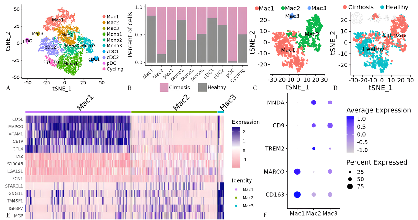
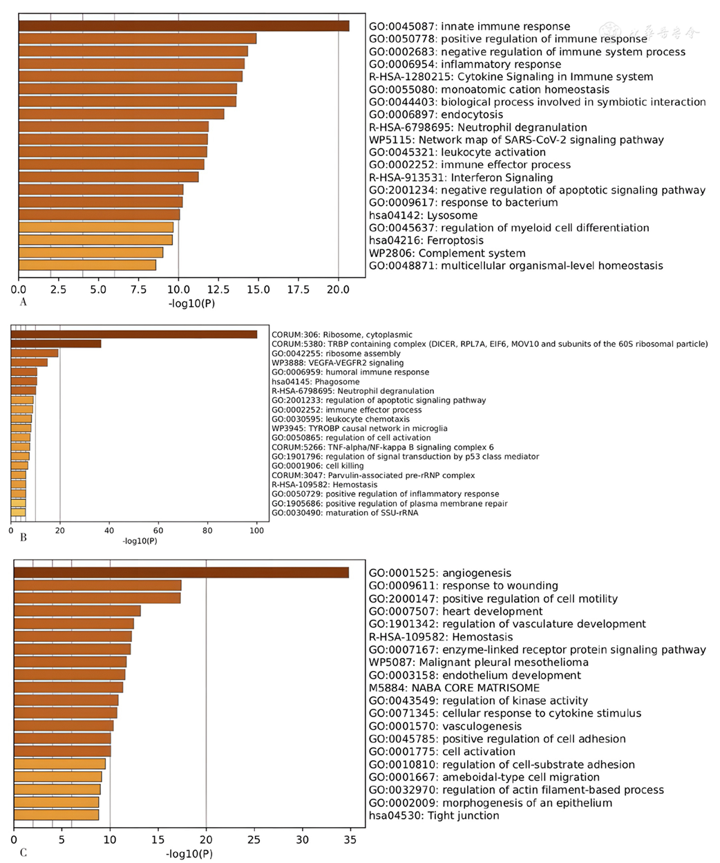
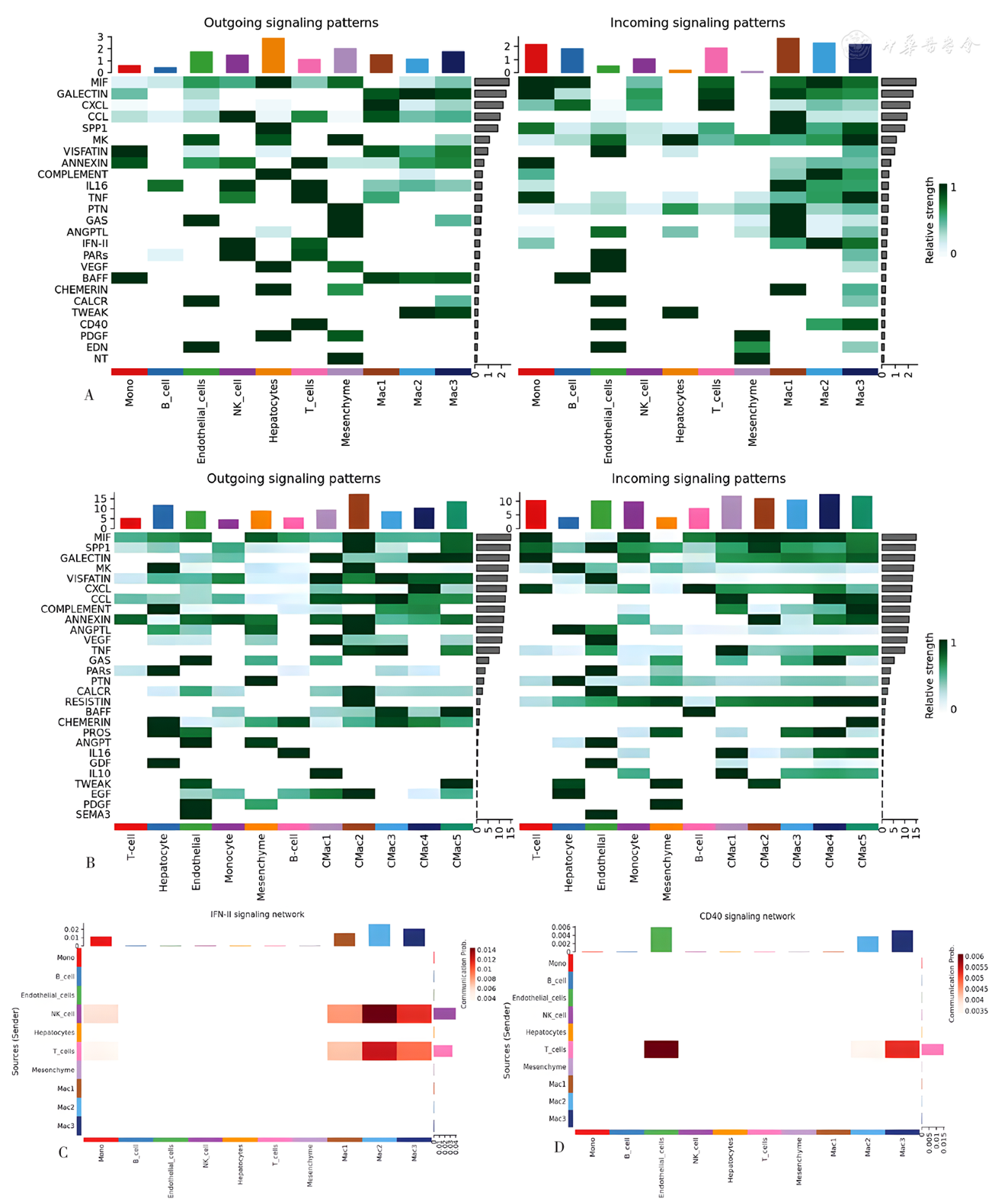
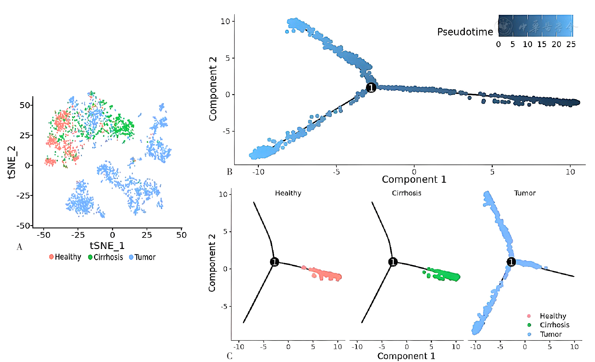
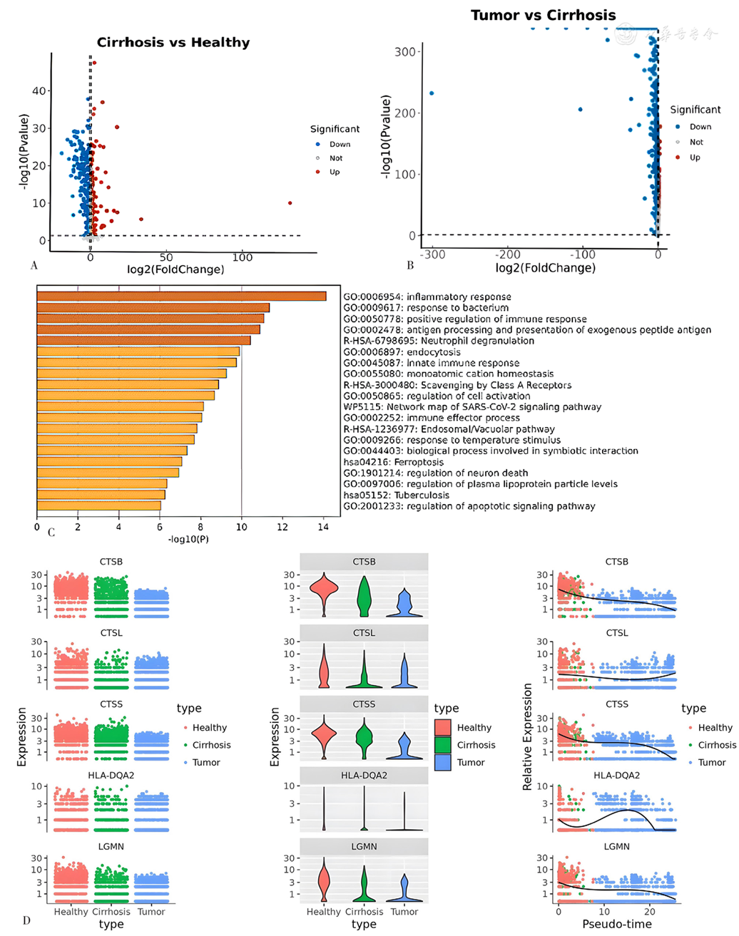
摘要: 背景 肝脏巨噬细胞在构建宿主防御机制及维持机体内环境稳定中发挥重要作用,也是参与肝脏损伤和修复的重要细胞成分。单核细胞来源的巨噬细胞在基因调控以及具体功能方面与肝脏固有巨噬细胞不尽相同。90%以上的原发性肝癌发生在肝硬化的基础上,巨噬细胞在肝硬化及肝癌疾病进展中的动态变化规律值得探讨。 目的 解析不同来源肝脏巨噬细胞的转录组学差异,分析巨噬细胞在肝硬化-肝癌疾病进展中的动态变化规律,探索预防肝硬化进展为肝癌的潜在策略。 方法 本研究通过从GEO数据库获取健康、肝硬化及肝癌组织的单细胞转录组学数据。健康及肝硬化数据来自GEO数据库GSE136103数据集,取自5例健康肝脏以及5例肝硬化肝脏的数据。肝癌数据来自GEO数据库GSE149614数据集,取自10例肝癌患者的数据。通过Seurat软件包分别对肝硬化及肝癌样本的数据进行聚类,鉴定各个细胞类型。将肝硬化样本中的3簇巨噬细胞亚群提取后,分析各个亚群前200个特异性表达基因,应用Metascape在线分析软件对各亚簇特异性表达基因进行功能分析。提取巨噬细胞亚群肝硬化特异性表达基因,通过KEGG功能分析探究巨噬细胞在肝硬化中的功能。将肝硬化以及肝癌单细胞转录组数据通过CellChat软件包进行细胞间相互作用分析,对比肝硬化与肝癌样本巨噬细胞的细胞通讯的差异。将健康对照、肝硬化以及肝癌三者不同来源的巨噬细胞通过Harmony软件包去批次效应,之后导入Monocle软件包进行伪时序分析,构建健康肝脏-肝硬化肝脏-肝癌巨噬细胞的演变轨迹。利用limma软件包找寻在健康肝脏-肝硬化肝脏-肝癌巨噬细胞的演变过程中连续上调以及下调的基因,并进行功能富集分析。 结果 对所有细胞进行无监督聚类,根据标记基因表达情况,共提取出3个巨噬细胞亚簇(分别为Mac1,Mac2和Mac3)。其中Mac1起源于组织驻留巨噬细胞(Kupffer细胞),Mac2以及Mac3起源于血液单核细胞,并且其数量在肝硬化组织中明显增多。在肝硬化组织中的Mac1表现了适应性免疫系统相关功能的上调,Mac2以及Mac3亚群均表现出吞噬体相关功能以及抗原提呈功能的下调。肝硬化与肝癌样本中巨噬细胞与其他类型细胞的通讯存在巨大的差异。某些细胞间通讯仅发生于肝硬化巨噬细胞中,这包括干扰素Ⅱ(IFN-Ⅱ)以及CD40等信号通路的细胞通讯。经过去批次效应的处理后,对健康肝脏、肝硬化肝脏以及肝癌巨噬细胞进行伪时序分析,结果提示三组数据存在特定的时序关系。本研究发现81个在该过程中连续下调的基因,然而未发现在健康肝脏-肝硬化肝脏-肝癌巨噬细胞演变过程中连续上调的基因。功能分析提示连续下调基因存在对细菌免疫反应的功能富集。 结论 肝硬化巨噬细胞可以分为3个亚群,其中Mac1来自肝脏固有Kupffer细胞,Mac2、Mac3来自血液单核细胞。肝硬化中诸多免疫相关细胞通讯例如IFN-Ⅱ以及CD40通路在肝癌中消失。健康肝脏-肝硬化肝脏-肝癌巨噬细胞演变过程存在对细菌免疫反应的持续下调,这可能加重了门脉高压造成的肠道菌群位移的危害。对于肝硬化患者,尽早地治疗门脉高压造成的肠漏,可能是重要的治疗策略。

关键词: 巨噬细胞, 肝硬化, 肝纤维化, 肝肿瘤, 单细胞转录组学测序, 细胞间相互作用

Abstract:

Background

Hepatic macrophages play a vital role in the defense mechanisms and maintaining the internal environment stability of body, and are also major cellular components involved in liver injury and repair. Macrophages derived from hematopoietic stem cells exhibit distinct gene regulation patterns compared to resident macrophages in the liver. More than 90% of primary liver cancer occurs on the basis of cirrhosis, and the dynamic changes of macrophages in the progression of cirrhosis to hepatocellular carcinoma are worth exploring.

Objective

To analyze the transcriptomic differences of hepatic macrophages originating from diverse sources, analyze the dynamic pattern of macrophage changes in liver cirrhosis and liver cancer progression, and explore potential strategies for preventing the progression of liver cancer.

Methods

In this study, single-cell transcriptomics data of healthy, cirrhotic and hepatocellular carcinoma (HCC) tissues were obtained from the Gene Expression Omnibus (GEO) database. The healthy and liver fibrosis data were obtained from the GSE136103 dataset of the GEO database, which included samples from five healthy liver tissues and five liver cirrhosis tissues. The HCC data were obtained from the GSE149614 dataset of the GEO database, which consisted of 21 samples from ten HCC patients. Utilizing the Seurat package, a clustering analysis was conducted on the transcriptomic data derived from liver fibrosis and HCC samples to identify distinct cell types. Notably, three distinctive clusters of macrophage subtypes were identified within the fibrosis samples, from which the top 200 marker genes were extracted. Metascape online analysis software was applied to functionally analyze each subcluster-specific expressed gene. Subgroup-specific expressed genes in liver fibrosis were extracted, and the function of macrophages in cirrhosis was explored by KEGG functional analysis. The CellChat software package was utilized to analyze intercellular interactions within liver fibrosis and HCC single-cell transcriptome data, differences in macrophage communication between cirrhosis and HCC samples were compared. Additionally, normal, fibrotic and cancerous macrophages were extracted, and batch effect correction was performed using the Harmony package. Subsequently, the Monocle package was employed for pseudo-time analysis to construct the developmental trajectory of macrophages spanning from a healthy state to fibrosis and eventually to the HCC microenvironment. The limma package was utilized to find genes that are continuously up-regulated and down-regulated during the evolution of macrophages from healthy state to cirrhotic state and finally to HCC, and functional enrichment analysis was performed.

Results

Unsupervised clustering was performed, and a total of three macrophage subclusters (designated as Mac1, Mac2, Mac3) were identified based on the expression patterns of marker genes. Mac1 originates from tissue-resident macrophages (Kupffer cells). Mac2 and Mac3 derived from blood monocytes and their numbers were significantly increased in cirrhotic tissue. Mac1 in cirrhotic tissue showed up-regulation of adaptive immune system-related functions. Mac2 and Mac3 subgroups show down-regulation of phagosome-related functions and antigen presentation functions. There were significant differences in communication between macrophages and other cell types in cirrhotic tissue and HCC tissue. Certain intercellular communication occurs only in cirrhotic macrophages, including cell communication of signaling pathways such as IFN-Ⅱ and CD40. After batch effect correction, pseudo-time series analysis was performed on macrophages from healthy liver, liver cirrhosis and HCC, the results suggest that there is a specific temporal relationship between the three groups of macrophages. This study identified 81 genes that were continuously down-regulated during the process, however, no genes were identified that were continuously up-regulated during the evolution of healthy-cirrhotic-HCC macrophage. Functional analysis suggested that the continuously down-regulated genes are functionally enriched for immune responses to bacteria.

Conclusion

Cirrhotic macrophages can be divided into three subgroups, of which Mac1 derived from liver-resident Kupffer cells and Mac2 and Mac3 derive from blood monocytes. Many immune-related cell communications in liver cirrhosis, such as IFN-Ⅱ and CD40 pathways, disappear in HCC. There is a continuous down-regulation of immune responses to bacteria in the evolution of healthy -cirrhotic-HCC macrophages, which may exacerbate the destructive effect of portal hypertension-induced gut microbiota displacement. For patients with liver cirrhosis, early treatment of portal hypertension-induced intestinal leakage (leaky gut) may be an important treatment strategy.

Key words: Macrophages, Liver cirrhosis, Liver fibrosis, Liver neoplasms, Single-cell transcriptomic sequencing, Intercellular interactions

中图分类号: