| [1] |
|
| [2] |
CHEUNG C Y, BIOUSSE V, KEANE P A,et al. Hypertensive eye disease[J]. Nat Rev Dis Primers, 2022, 8:14. DOI: 10.1038/s41572-022-00342-0.
|
| [3] |
KLEIN R, KLEIN B E, MOSS S E. The relation of systemic hypertension to changes in the retinal vasculature:the Beaver Dam Eye Study[J]. Trans Am Ophthalmol Soc,1997,95:329-348;discussion 348-350.
|
| [4] |
AKRAM M U, AKBAR S, HASSAN T,et al. Data on fundus images for vessels segmentation,detection of hypertensive retinopathy,diabetic retinopathy and papilledema[J]. Data Brief, 2020, 29:105282. DOI: 10.1016/j.dib.2020.105282.
|
| [5] |
FENG X, WANG H W, KONG Y Y,et al. Diagnosis of chronic stage of hypertensive retinopathy based on spectral domain optical coherence tomography[J]. J Clin Hypertens, 2020, 22(7):1247-1252. DOI: 10.1111/jch.13935.
|
| [6] |
LEE W H, LEE M W, LIM H B,et al. Retinal nerve fibre layer/ganglion cell-inner plexiform layer thickness ratio in patients with systemic hypertension[J]. Acta Ophthalmol, 2022, 100(1):e150-156. DOI: 10.1111/aos.14884.
|
| [7] |
WHELTON P K, WILLIAMS B. The 2018 European society of cardiology/european society of hypertension and 2017 American college of cardiology/american heart association blood pressure guidelines:more similar than different[J]. JAMA, 2018, 320(17):1749-1750. DOI: 10.1001/jama.2018.16755.
|
| [8] |
WALSH J B. Hypertensive retinopathy. description,classification,and prognosis[J]. Ophthalmology,1982,89(10):1127-1131.
|
| [9] |
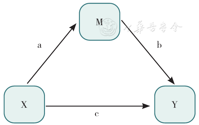
BARON R M, KENNY D A. The moderator-mediator variable distinction in social psychological research:conceptual,strategic,and statistical considerations[J]. J Pers Soc Psychol, 1986, 51(6):1173-1182. DOI: 10.1037//0022-3514.51.6.1173.
|
| [10] |
VALERI L, VANDERWEELE T J. Mediation analysis allowing for exposure-mediator interactions and causal interpretation:theoretical assumptions and implementation with SAS and SPSS macros[J]. Psychol Methods, 2013, 18(2):137-150. DOI: 10.1037/a0031034.
|
| [11] |
CÁCERES TOLEDO M CTO, CORDIÉS JACKSON L. Hipertensión arterial y retinopatía hipertensiva:Su comportamiento en un área de salud [J]. Revista Cubana de Medicina,2000,39(4):210-216.
|
| [12] |
WONG T Y, MITCHELL P. Hypertensive retinopathy[J]. N Engl J Med, 2004, 351(22):2310-2317. DOI: 10.1056/NEJMra032865.
|
| [13] |
DZIEDZIAK J, ZALESKA-ŻMIJEWSKA A, SZAFLIK J P,et al. Impact of arterial hypertension on the eye:a review of the pathogenesis,diagnostic methods,and treatment of hypertensive retinopathy[J]. Med Sci Monit, 2022, 28:e935135. DOI: 10.12659/MSM.935135.
|
| [14] |
AKAY F, GÜNDOĞAN F C, YOLCU U,et al. Retinal structural changes in systemic arterial hypertension:an OCT study[J]. Eur J Ophthalmol, 2016, 26(5):436-441. DOI: 10.5301/ejo.5000740.
|
| [15] |
LEE W H, LEE M W, LIM H B,et al. Longitudinal changes in the thickness of the ganglion cell-inner plexiform layer in patients with hypertension:a 4-year prospective observational study[J]. Acta Ophthalmol, 2020, 98(4):e479-486. DOI: 10.1111/aos.14291.
|
| [16] |
IADECOLA C. The neurovascular unit coming of age:a journey through neurovascular coupling in health and disease[J]. Neuron, 2017, 96(1):17-42. DOI: 10.1016/j.neuron.2017.07.030.
|
| [17] |
|
| [18] |
PRESA J L, SARAVIA F, BAGI Z,et al. Vasculo-neuronal coupling and neurovascular coupling at the neurovascular unit:impact of hypertension[J]. Front Physiol, 2020, 11:584135. DOI: 10.3389/fphys.2020.584135.
|
| [19] |
CÁRDENAS L T G, MEJÍAS L C, PEREA L P,et al. The Family Doctor and Nurse Program:development of the health care model in CubaO Programa de Médicos e Enfermeiras da Família:desenvolvimento do modelo de saúde em Cuba[J]. Rev Panam Salud Publica, 2018, 42:e31. DOI: 10.26633/RPSP.2018.31.
|
| [20] |
吴述银,谢云涛,刘露,等. 社区居民对医联体专科医生参与家庭医生签约服务的认知分析[J]. 南京医科大学学报:社会科学版,2022,22(6):591-596.
|
| [21] |
|

 )
)