中国全科医学 ›› 2022, Vol. 25 ›› Issue (15): 1869-1874.DOI: 10.12114/j.issn.1007-9572.2022.02.004

• 论著 • 上一篇    下一篇

原发性甲状腺淋巴瘤的超声特征研究

欧阳向柳1, 高蓓1, 王艳滨1, 刘丽云2, 顾程3, 郑立春3,*()   

  1. 1. 063000 河北省唐山市工人医院超声科
    2. 063000 河北省唐山市工人医院病理科
    3. 063000 河北省唐山市工人医院核医学科
  • 收稿日期:2021-11-18 修回日期:2022-01-10 出版日期:2022-03-17 发布日期:2022-04-07
  • 通讯作者: 郑立春
  • 欧阳向柳,高蓓,王艳滨,等.原发性甲状腺淋巴瘤的超声特征研究[J].中国全科医学,2022,25(15):1869-1874. [www.chinagp.net]
    作者贡献:欧阳向柳提出研究方向,确定主要研究目标,负责文献的收集,文章的构思与设计,撰写论文初稿;高蓓、王艳滨、刘丽云负责文章的构思,回顾性整理病例资料,整理图表;顾程对论文初稿及英文摘要进行修订;郑立春对论文终稿进行审校,负责文章的质量控制。
  • 基金资助:
    河北省省级科技计划基金资助项目(182777145); 河北省医学科学研究课题计划基金资助项目(20201517)

Ultrasonographic Analysis of Primary Thyroid Lymphoma

Xiangliu OUYANG1, Bei GAO1, Yanbin WANG1, Liyun LIU2, Cheng GU3, Lichun ZHENG3,*()   

  1. 1. Department of Ultrasound, Tangshan Gongren Hospital, Tangshan 063000, China
    2. Department of Pathology, Tangshan Gongren Hospital, Tangshan 063000, China
    3. Department of Nuclear Medicine, Tangshan Gongren Hospital, Tangshan 063000, China
  • Received:2021-11-18 Revised:2022-01-10 Published:2022-03-17 Online:2022-04-07
  • Contact: Lichun ZHENG
  • About author:
    OUYANG X L, GAO B, WANG Y B, et al. Ultrasonographic analysis of primary thyroid lymphoma[J]. Chinese General Practice, 2022, 25 (15) : 1869-1874.

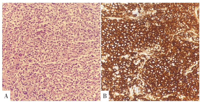
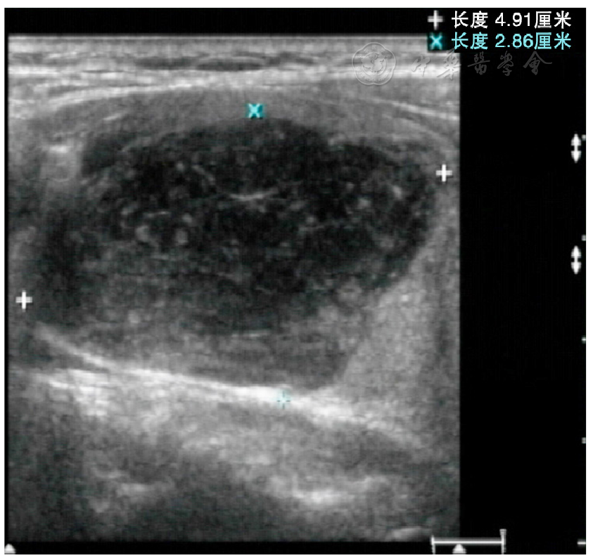
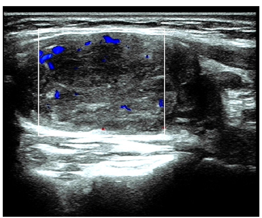
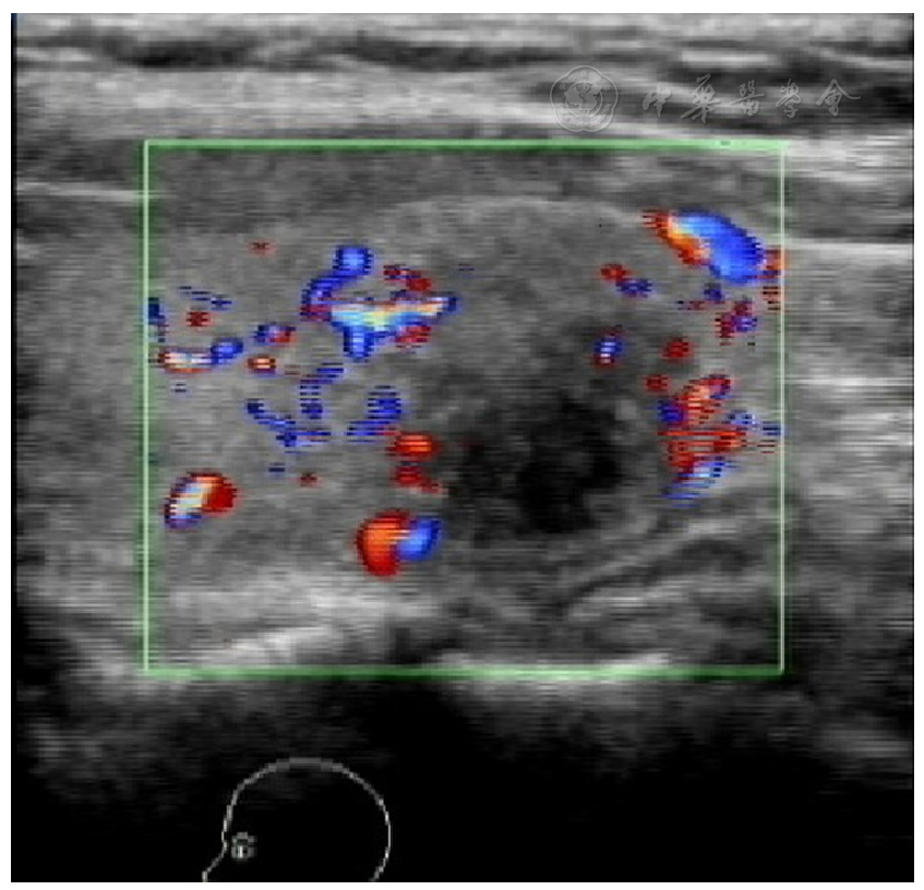
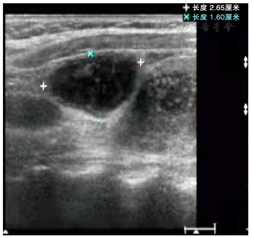
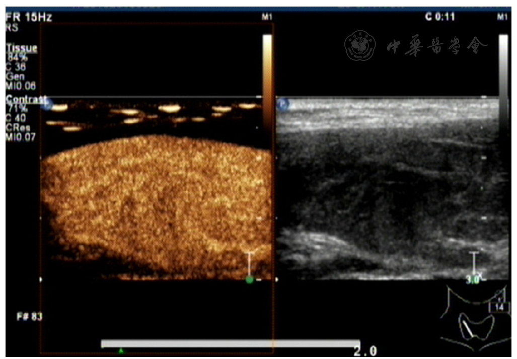
摘要: 背景 原发性甲状腺淋巴瘤(PTL)是较为罕见的结外型淋巴瘤类型,临床特征不典型,超声特征不明显,经常被误诊和漏诊。 目的 总结分析PTL的二维超声及超声造影声像图特征表现。 方法 回顾性分析2012年12月至2020年12月唐山市工人医院收治的16例经病理证实的PTL患者(PTL组)的超声声像图特征,并与同期收治的16例甲状腺癌患者(甲状腺癌组)的超声声像图进行对比分析。 结果 PTL组病理结果:10例为弥漫性大B细胞淋巴瘤、4例为黏膜相关淋巴组织结外边缘区B细胞淋巴瘤、1例为滤泡性淋巴瘤、1例为伯基特淋巴瘤。甲状腺癌组病理结果:11例为乳头状癌、4例为滤泡癌、1例为髓样癌。PTL组甲状腺体积增大率(13/16)高于甲状腺癌组(5/16)(χ2=8.127,P<0.05),病灶后方回声增强率(14/16)高于甲状腺癌组(3/16)(χ2=15.184,P<0.05),钙化率(0例)低于甲状腺癌组(10/16)(χ2=14.545,P<0.05)。PTL组患者中13例病灶形态较规则、边界清晰,甲状腺癌组中12例病灶形态不规则、边界不清晰,两组的病灶形态和边界特征比较,差异有统计学意义(χ2=10.165,P<0.05)。PTL组(7/16)和甲状腺癌组(3/16)病灶血流信号增多率比较,差异无统计学意义(χ2=3.327,P>0.05)。PTL组(8/16)和甲状腺癌组(11/16)颈部淋巴结转移率比较,差异无统计学意义(χ2=1.166,P>0.05)。超声造影PTL动脉期及静脉期多呈高增强,甲状腺癌动脉期及静脉期多呈低增强。 结论 PTL的超声表现有一定的特征性,甲状腺体积增大,伴弥漫性极低回声病灶,无钙化,病灶形态规则、边界清晰,后方回声增强,造影动脉期及静脉期呈高增强时应考虑为PTL。

关键词: 甲状腺, 淋巴瘤, 超声检查, 超声造影, 甲状腺肿瘤

Abstract:

Background

Primary thyroid lymphoma (PTL) is a rare type of extranodal lymphoma, which is prone to misdiagnosis and missed diagnosis due to atypical clinical and ultrasonic features.

Objective

To summarize and analyze the features of 2-D ultrasonography and contrast-enhanced ultrasonography in PTL.

Methods

A retrospective, comparative analysis was conducted on ultrasonographic features of 16 patients with pathologically confirmed PTL (PTL group) and 16 thyroid cancer patients (thyroid cancer group) recruited from Tangshan Gongren Hospital during December 2012 to December 2020.

Results

Pathological results of PTL group: 10 cases were diffuse large B-cell lymphoma, 4 cases were mucous-associated lymphoid tissue extranodular marginal zone B-cell lymphoma, 1 case was follicular lymphoma, and 1 case was Burkitt lymphoma. Pathological results of thyroid cancer group: 11 cases were papillary carcinoma, 4 cases were follicular carcinoma and 1 case was medullary carcinoma. The rate of thyroid enlargement in PTL group was higher than that in thyroid cancer group (13/16 vs 5/16) (χ2=8.127, P<0.05) . PTL group also had higher rate of posterior echo enhancement (14/16 vs 3/16) (χ2=15.184, P<0.05) . Besides that, PTL group had lower calcification rate (0) than thyroid cancer group did (10/16) (χ2=14.545, P<0.05) . PTL and thyroid cancer groups had statistically differences in lesion morphology and margins (χ2=10.165, P<0.05) as 13 PTL cases had lesions with regular morphology and well-defined margins, and 12 thyroid cancer cases had lesions with irregular morphology and ill-defined margins. The rate of increased blood flow signals within the lesions showed no significant difference between PTL group and thyroid cancer group (7/16 vs 3/16) (χ2=3.327, P>0.05) . PTL and thyroid cancer groups also had no statistically significant difference in the rate of cervical lymph node metastases (8/16 vs 11/16) (χ2=1.166, P>0.05) . Contrast-enhanced ultrasonography showed high enhancement during arterial and venous phases in PTL, and low enhancement at arterial and venous phases in thyroid cancer.

Conclusion

PTL has specific ultrasonic manifestations. A diagnosis of PTL could be made when a patient manifests as thyroid enlargement, diffuse hypoechoic, non-calcified, regular-shaped lesions with well-defined margins and posterior echo enhancement, and high enhancement at arterial and venous phases.

Key words: Thyroid, Lymphoma, Ultrasonography, Contrast-enhanced ultrasonography, Thyroid neoplasms