中国全科医学 ›› 2022, Vol. 25 ›› Issue (29): 3691-3697.DOI: 10.12114/j.issn.1007-9572.2022.0300

• 论著·诊疗提示 • 上一篇    下一篇

胎儿动静脉畸形的产前超声诊断研究

谭筱檀, 袁红霞*()   

  1. 410007 湖南省长沙市妇幼保健院超声科
  • 收稿日期:2022-02-15 修回日期:2022-05-06 出版日期:2022-10-15 发布日期:2022-07-07
  • 通讯作者: 袁红霞
  • 谭筱檀,袁红霞.胎儿动静脉畸形的产前超声诊断研究[J].中国全科医学,2022,25(29):3691-3697. [www.chinagp.net]
    作者贡献:袁红霞提出将胎儿动静脉畸形病例进行归纳总结的构想,为撰写论文提供病例报告及影像资料,负责部分病例的超声检查,对疑难病例进行会诊并给出产前诊断;谭筱檀进行研究命题的设计,提出借鉴YAKES分型对胎儿动静脉畸形进行分类研究,负责部分病例的超声检查,收集病例详细的临床资料,论文的撰写以及最终版本修订,对论文整体负责。
  • 基金资助:
    湖南省卫生健康委科研计划项目资助(202209023567)

Prenatal Ultrasound Diagnosis of Fetal Arteriovenous Malformations

Xiaotan TAN, Hongxia YUAN*()   

  1. Department of Ultrasound, Changsha Hospital for Maternal and Child Health Care, Changsha 410007, China
  • Received:2022-02-15 Revised:2022-05-06 Published:2022-10-15 Online:2022-07-07
  • Contact: Hongxia YUAN
  • About author:
    TAN X T, YUAN H X. Prenatal ultrasound diagnosis of fetal arteriovenous malformations[J]. Chinese General Practice, 2022, 25 (29) : 3691-3697.

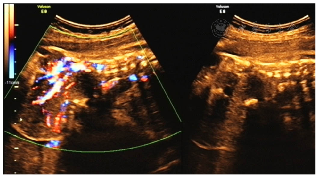
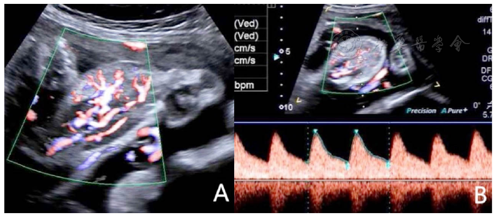
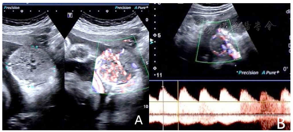
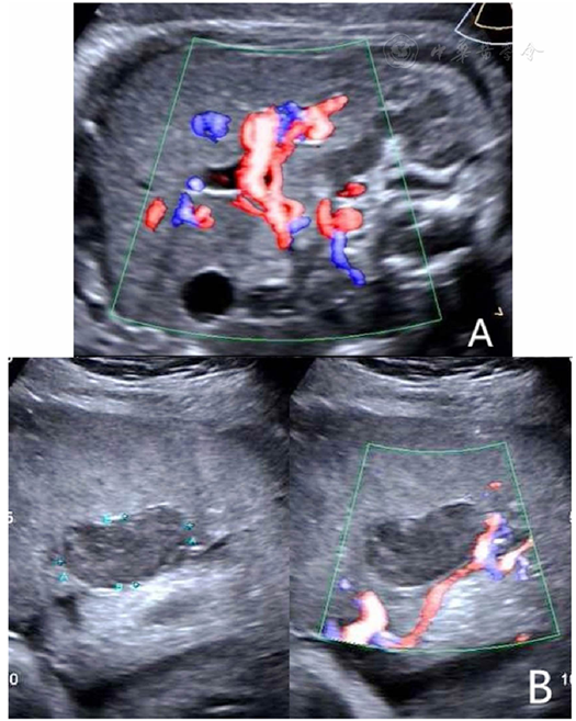
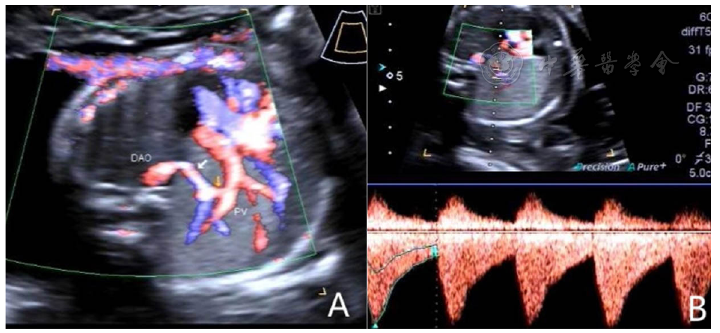
摘要: 背景 胎儿动静脉畸形可发生在全身各个部位,主要依靠彩色多普勒超声进行诊断,而关于产前超声诊断动静脉畸形类型的探讨国内外鲜有报道。 目的 探讨产前超声诊断胎儿动静脉畸形的价值。 方法 选取2013年8月至2021年8月在长沙市妇幼保健院超声科行产前超声筛查胎儿动静脉畸形的孕妇为研究对象,采用彩色多普勒超声诊断仪进行胎儿系统超声筛查,发现胎儿动静脉畸形病灶时,描述其位置、大小、形态、内部回声。分析不同部位动静脉畸形的超声特征及动静脉畸形分型,并追踪随访结果。 结果 本研究中产前超声筛查发现胎儿动静脉畸形病例16例,其中6例病灶位于胎儿骶尾部(诊断为骶尾部畸胎瘤),4例位于胎儿颅内(包括3例颅内蔓状血管瘤合并Galen静脉瘤、1例Galen静脉瘤),3例位于胎儿肢体、软组织(包括1例Parkes-Weber综合征、2例软组织血管瘤),2例位于胎儿肝脏(诊断为肝动静脉瘘),1例位于胎儿肺部(诊断为隔离肺并肺动静脉瘘)。动静脉畸形分型:单纯Ⅱ型10例(10/16),Ⅱ型和Ⅲa型同时存在3例(3/16),单纯Ⅰ型1例(1/16),单纯Ⅳ型1例(1/16),单纯Ⅲa型1例(1/16)。随访显示,10例引产,3例失访,2例足月出生后行手术治疗,1例死胎。 结论 胎儿动静脉畸形的预后与病变部位、病灶内血流分布情况关系密切。产前超声检查可发现胎儿有无动静脉畸形病灶,对其诊断、预后判断具有重要价值。

关键词: 动静脉畸形, 产前诊断, 超声检查,产前, 胎儿, 超声检查,多普勒,彩色

Abstract:

Background

Fetal arteriovenous malformations may occur in all parts of the body and are mainly diagnosed by color Doppler ultrasound. However, there are few reports at home and abroad on the types of arteriovenous shunts explored by prenatal ultrasound.

Objective

To investigate the diagnosis value of prenatal ultrasonography in fetal arteriovenous malformation.

Methods

Pregnant women with fetal arteriovenous malformations who underwent prenatal ultrasound screening in the Department of Ultrasound, Changsha Hospital for Maternal and Child Health Care from August 2013 to August 2021 were recruited. Color Doppler ultrasonography was used for fetal systematical ultrasound screening. The location, size, shape, and internal echoes of fetal arteriovenous malformation lesions were detected and described. The ultrasonographic features and types of arteriovenous malformations in different parts of patients were analyzed, and the results of follow-up were tracked.

Results

Prenatal ultrasound screening in this study identified sixteen cases with fetal arteriovenous malformations, of which six lesions were located in the fetal sacrococcygeal region (diagnosed as sacrococcygeal teratoma) , four were located in the fetal intracranial region (including three intracranial cranberg hemangiomas with Galen's vein tumors, one Galen's vein tumor) , three were located in the fetal limbs, soft tissues (including one Parkes Weber syndrome, two soft tissue hemangiomas) , two were located in fetal liver (diagnosed as hepatic arteriovenous fistula) and one was located in fetal lung (diagnosed as isolated lung with pulmonary arteriovenous fistula) . Arteriovenous malformation typing: 10 patients had simple type Ⅱ (10/16) , 3 patients had both type Ⅱ and type Ⅲa (3/16) , 1 patient had type Ⅰ (1/16) , 1 patient had type Ⅳ (1/16) , 1 patient had simple type Ⅲa (1/16) . Follow up showed that 10 induced labor, 3 were lost to follow-up, 2 were born at term and treated surgically, and 1 was stillborn.

Conclusion

The prognosis of fetal arteriovenous malformation is closely related to the location of lesion and blood flow distribution within the lesion. Prenatal ultrasound can detect a fetus with or without a focus of arteriovenous malformation, which is valuable for diagnosis and prognosis, and can help to detect the potential threat caused by arteriovenous malformation to the fetus.

Key words: Arteriovenous malformations, Prenatal diagnosis, Ultrasonography, prenatal, Fetus, Ultrasonography, doppler, color