
Page 114 - 中国全科医学2022-14
P. 114
http://www.chinagp.net E-mail:zgqkyx@chinagp.net.cn ·1775·
部急性损伤或肉芽肿性病变。以下列举部分药物引起的肺组 他肺损害(ILD 的其他亚型、非 ILD 表型)和其他原因导致
织病理表现(图 5~7)。 的 OP。
4 DIOP 的鉴别诊断 4.1 其他亚型的 DIILD 药物也可引起其他类型 ILD,ILD
DIOP 的鉴别主要分为两个思路,分别为药物诱导的其 在症状上均可表现为干咳和呼吸困难,主要有 HP、NSIP、
急 性 间 质 性 肺 炎 - 成 人 呼 吸 窘 迫 综 合 征(acute interstitial
pneumonia-adult respiratory distress syndrome,AIP-ARDS)、
结节样肉芽肿表型 [36] ,各类型之间的鉴别需从临床表现、
影像学及病理检查多方面综合考量,建议通过多学科讨论
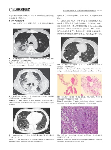
图 1 阿扎胞苷使用 21 d 后 CT:邻近胸膜的实变影伴周围磨玻璃影
及支气管充气征(引自文献[47])
Figure 1 CT after 21 days of azacitidine use:consolidation of adjacent
pleura with surrounding ground-glass shadow and bronchial inflation sign 图 4 另一患者 nivolumab 使用 4 个月后 CT:外周及胸膜下为主的多
发实变影(引自文献[36])
Figure 4 Another patient's CT after using nivolumab for 4 months:
multiple consolidations predominantly in the periphery and under the pleura
图 2 依维莫司使用 4 周后 CT:支气管中心性实变伴双侧广泛轻微网
图 5 阿扎胞苷,CT 引导下穿刺活检病理:机化性肺炎,慢性非特
状影及间隔增厚(引自文献[25])
异性炎症伴巨噬细胞(引自文献[15])
Figure 2 CT after 4 weeks of everolimus use:central bronchial
Figure 5 Azacitidine,CT-guided needle biopsy pathology:organizing
consolidation with bilateral extensive slight reticular shadow and septal
pneumonia,chronic non-specific inflammation with macrophages
thickening
图3 Nivolumab使用4个月后CT:中下肺外周及胸膜下多发实变影(引 图 6 依维莫司,肺楔叶切除组织病理:机化性肺炎,伴急性肺损伤
自文献[36]) 的特征(引自文献[25])
Figure 3 CT after nivolumab use for 4 months:multiple consolidations in Figure 6 Everolimus,pulmonary cuneiectomy histopathology:organizing
the periphery of the middle and lower lung and subpleural pneumonia,with features of acute lung injury