Chinese General Practice ›› 2026, Vol. 29 ›› Issue (29): 4281-4287.DOI: 10.12114/j.issn.1007-9572.2025.0130

• Article • Previous Articles     Next Articles

Automatic Segmentation of Colorectal Cancer Lesions in Abdominal CT Images Using a Deep Learning Network Based on Fused 3D U-Net and Transformer: a Multicenter and Multidevice Study


  1. 1. School of Biomedical Engineering, Southern Medical University, Guangzhou 510515, China
    2. Department of Radiology, Taizhou Hospital of Zhejiang Province, Linhai 317000, China
    3. Department of Radiology, Guangdong Provincial Traditional Chinese Medicine Hospital/The Second Affiliated Hospital of Guangzhou University of Chinese Medicine, Guangzhou 510120, China
    4. Department of Radiology, Nanfang Hospital, Southern Medical University, Guangzhou 510515, China
    5. Department of Radiology, Sun Yat-Sen Memorial Hospital, Sun Yat-sen University, Guangzhou 510235, China
  • Received:2024-03-14 Revised:2025-06-18 Published:2026-10-15 Online:2026-09-02
  • Contact: CHEN Weicui

基于融合三维U-Net与Transformer的深度学习网络对腹部CT图像中结直肠癌病灶的自动分割:一项多中心、多设备研究


  1. 1.510515 广东省广州市,南方医科大学生物医学工程学院
    2.317000 浙江省临海市,浙江省台州医院放射科
    3.510120 广东省广州市,广东省中医院 广州中医药大学第二附属医院影像科
    4.510515 广东省广州市,南方医科大学南方医院影像科
    5.510235 广东省广州市,中山大学孙逸仙纪念医院影像科
  • 通讯作者: 陈维翠
  • 作者简介:

    作者贡献:

    黄牧、张道春及陈维翠提出研究思路,设计研究方案;黄牧负责构建及验证深度学习模型;阳维、钟丽明提供方法学支持;袁文静、贾子琪负责数据收集及整理;谭相良、段小慧负责提供外部验证数据;刘岘负责统计学分析;陈维翠负责最终版本修订,对论文负责。

  • 基金资助:
    国家自然科学基金资助项目(82202259); 广东省中医院第十三届朝阳人才项目(ZY2022YL05)

Abstract:

Background

The application of deep learning in medical imaging faces challenges such as time-consuming and labor-intensive data annotation, which hinders their clinical translation efficiency.

Objective

To investigate the feasibility and efficacy of a deep learning network (TransUNet-Cascade) that integrates 3D U-Net and Transformer for the automatic segmentation of colorectal cancer (CRC) lesions in abdominal CT images.

Methods

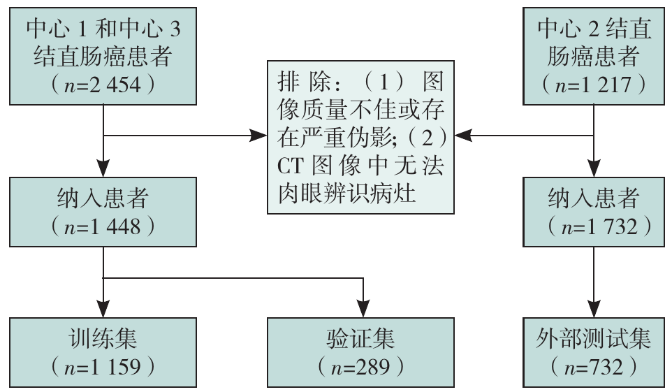
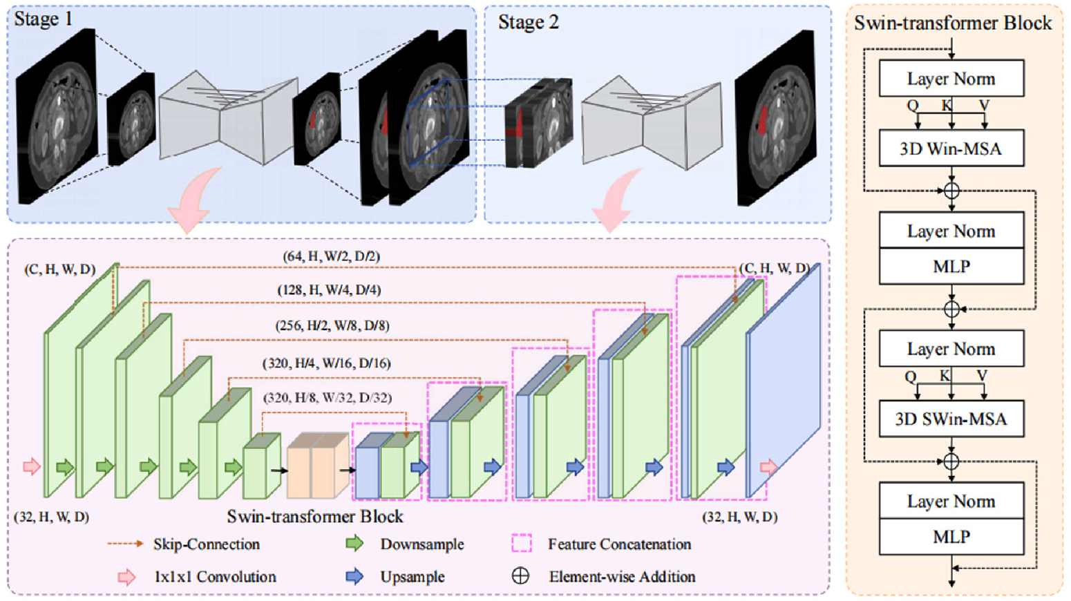
A retrospective analysis was conducted on contrast-enhanced abdominal CT images from 2 180 CRC patients at Guangdong Provincial Traditional Chinese Medicine Hospital (Center 1), Nanfang Hospital, Southern Medical University (Center 2), and Sun Yat-sen Memorial Hospital, Sun Yat-sen University (Center 3) between January 2018 and May 2023. The dataset was divided into a training set (n=1 159), a validation set (n=289), and an external test set (n=732) using weighted random sampling. A cascaded network framework (TransUNet-Cascade) based on 3D U-Net and Swin Transformer was proposed, optimizing segmentation accuracy through a multi-stage learning strategy. Model performance was evaluated using manual annotations as the gold standard, with metrics including Dice similarity coefficient (DSC), F1 score, 95% Hausdorff distance (HD95), intersection over union (IoU), precision (PRE), and recall rate (REC). This study selected the 3D no new U-Net (3D nnU-Net) as the baseline model for comparison. Under the same dataset and evaluation criteria, a systematic training and performance comparison was conducted with the proposed TransUNet-Cascade network to comprehensively verify its effectiveness in the CRC segmentation task.

Results

In the external test set, the performance of the two deep learning models based on the AP CT images was superior to that based on the VP CT images. TransUNet-Cascade achieved average DSC, F1 score, HD95, IoU, PRE, and REC values of 0.740, 0.839, 34.084, 0.656, 0.737, and 0.767, respectively, which were overall superior to 3D nnU-Net (average DSC, F1 score, HD95, IoU, PRE, and REC values of 0.724, 0.838, 35.954, 0.642, 0.730, and 0.744, respectively). The model demonstrated the best segmentation performance for right-sided colon cancer (DSC=0.784), while segmentation of rectal cancer was relatively less effective (DSC=0.622).

Conclusion

By combining the strengths of convolutional neural networks and Transformers, TransUNet-Cascade improves the accuracy of automatic CRC lesion segmentation, demonstrating potential for clinical application.

Key words: Colorectal neoplasms, Tomography, X-ray computed, Deep learning, Artificial intelligence, Self-attention mechanism, Convolutional neural network

摘要:

背景

深度学习在医学图像中的应用面临数据标注耗时、费力等挑战,制约了其临床转化效率。

目的

探讨一种融合三维U-Net(3D U-Net)与Transformer的深度学习网络(TransUNet-Cascade)在腹部CT图像中自动分割结直肠癌(CRC)病灶的可行性与效能。

方法

回顾性纳入2018年1月—2023年5月来自广东省中医院(中心1)、南方医科大学南方医院(中心2)及中山大学孙逸仙纪念医院(中心3)的2 180例CRC患者腹部CT增强图像,采用分层加权随机抽样法划分为训练集(n=1 159)、验证集(n=289)和外部测试集(n=732)。本研究提出一种新型深度学习网络模型——TransUNet-Cascade,通过多阶段学习策略优化分割精度。以手动标注为基准,采用Dice相似系数(DSC)、F1值、95%豪斯多夫距离(HD95)、交互比(IoU)、精确度(PRE)以及召回率(REC)评估模型性能。本研究选取三维no new U-Net(3D nnU-Net)作为对比基线模型,在统一的数据集和评估标准下,与所提出的TransUNet-Cascade网络进行系统训练与性能对比,以全面验证其在CRC分割任务中的有效性。

结果

在独立的外部测试集中,两种深度学习网络基于动脉期图像的分割效能总体优于静脉期图像;TransUNet-Cascade动脉期平均DSC、F1值、HD95、IoU、PRE及REC分别为0.740、0.839、34.084、0.656、0.737及0.767,总体优于3D nnU-Net(平均DSC、F1值、HD95、IoU、PRE及REC分别为0.724、0.838、35.954、0.642、0.730及0.744)。模型对右半结肠癌的分割效果最佳(DSC=0.784),直肠癌分割效果相对较差(DSC=0.622)。

结论

TransUNet-Cascade通过结合卷积神经网络与Transformer的优势,提升了CRC病灶的自动分割精度,具有一定的临床应用潜力。

关键词: 结直肠肿瘤, 体层摄影术,X线计算机, 深度学习, 人工智能, 自注意力机制, 卷积神经网络

CLC Number: