中国全科医学 ›› 2022, Vol. 25 ›› Issue (36): 4567-4572.DOI: 10.12114/j.issn.1007-9572.2022.0411

• 诊疗分析 • 上一篇    下一篇

巨大胎盘类血池样病变超声征象与临床诊断分析

玄英华, 王莉*(), 黄瑞贞, 吴青青   

  1. 100026 北京市,首都医科大学附属北京妇产医院 北京妇幼保健院超声科
  • 收稿日期:2022-03-18 修回日期:2022-07-03 出版日期:2022-12-20 发布日期:2022-07-21
  • 通讯作者: 王莉
  • 玄英华,王莉,黄瑞贞,等.巨大胎盘类血池样病变超声征象与临床诊断分析[J].中国全科医学,2022,25(36):4567-4572. [www.chinagp.net]
    作者贡献:玄英华提出研究思路,负责收集病例资料、随访病例、查阅文献以及起草论文;王莉负责研究设计、收集病例资料、修改论文,负责最终版本修订,对论文负责;黄瑞贞负责收集病例资料;吴青青为论文撰写提供建议。

Ultrasonographic Findings and Clinical Diagnosis of Large Placental Lake-like Lesions: Clinical Analysis of Six Cases

XUAN Yinghua, WANG Li*(), HUANG Ruizhen, WU Qingqing   

  1. Department of Ultrasound, Beijing Obstetrics and Gynecology Hospital, Capital Medical University/Beijing Maternal and Child Health Care Hospital, Beijing 100026, China
  • Received:2022-03-18 Revised:2022-07-03 Published:2022-12-20 Online:2022-07-21
  • Contact: WANG Li
  • About author:
    XUAN Y H, WANG L, HUANG R Z, et al. Ultrasonographic findings and clinical diagnosis of large placental lake-like lesions: clinical analysis of six cases [J] . Chinese General Practice, 2022, 25 (36) : 4567-4572.

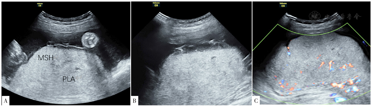
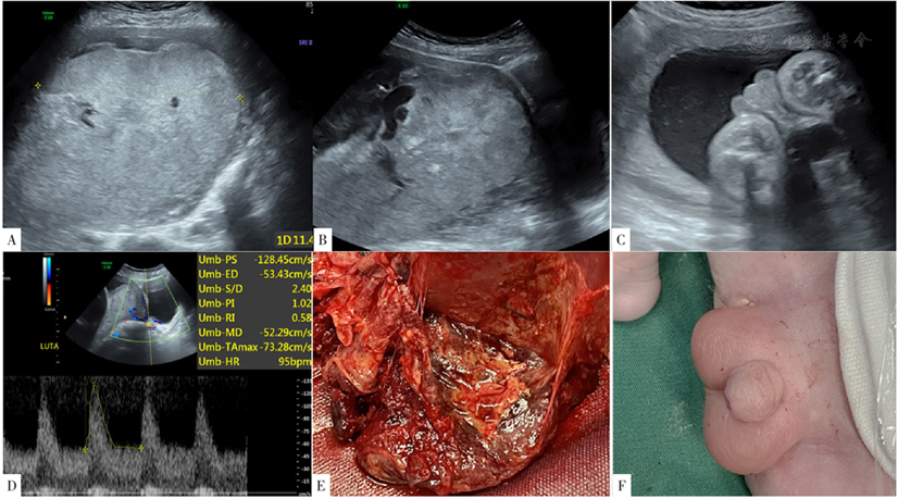
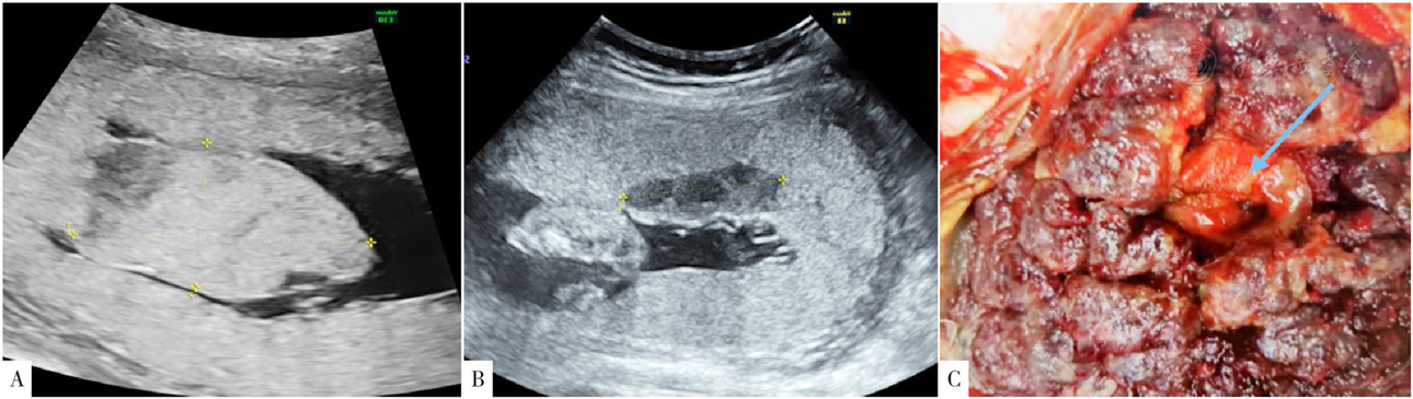
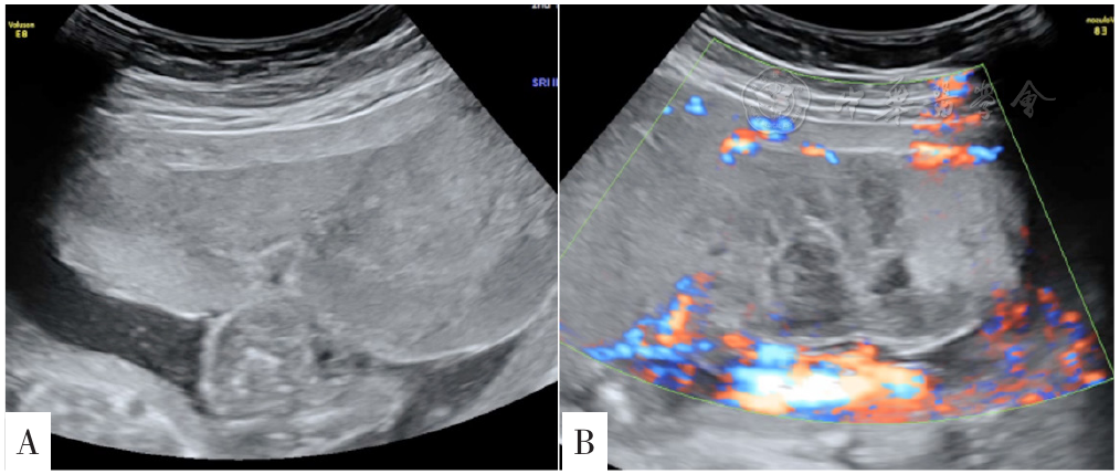
摘要: 目的 探讨不同巨大胎盘类血池样病变的产前超声特征及产前诊断意义。 方法 回顾性分析2016年2月至2021年12月在首都医科大学附属北京妇产医院进行超声检查时发现巨大胎盘类血池样病变的6例患者的临床资料,分析其一般资料、超声特征、临床诊断及妊娠结局。以最大径线>5 cm作为巨大胎盘类血池样病变的定义。 结果 6例患者均为单胎妊娠,年龄27~33岁,首次发现巨大胎盘类血池样异常回声的孕周为18+2~31+6周。3例(病例1、病例2及病例3)类血池样异常回声与胎盘组织有分界,分别于妊娠33+5周、29+1周及32周行剖宫产,3例新生儿均存在尿道下裂畸形,临床诊断均为胎盘绒毛膜板下出血;1例(病例4)类血池样病变位于胎盘子面,与胎盘组织有分界,突向羊膜腔,动态观察可以消失,妊娠期超声监测胎儿生长符合孕周,妊娠后期血池范围明显减小,临床诊断为巨大胎盘血池;2例(病例5和病例6)类血池样病变为弥漫性病变,累及大部分胎盘,正常胎盘组织减少,胎儿均为早发型生长受限(各经线<1%、腹围为2.3%),脐动脉舒张末期血流均消失,孕妇子宫动脉舒张早期出现切迹(双侧、单侧);病例5于妊娠23周终止妊娠,病例6并发重度子痫前期,于妊娠27+4周终止妊娠,临床诊断均为胎盘灌注不良合并早发型胎儿生长受限。 结论 妊娠期胎盘类血池样异常回声根据其不同形成原因,有各自的超声特征,产前可以进行鉴别,及时诊断有助于提示严重胎盘灌注不良及绒毛膜板下出血病例产前严密监测胎儿情况,减少不良妊娠结局的发生。

关键词: 胎盘, 血池, 胎儿宫内监测, 胎儿宫内生长受限, 早发型, 尿道下裂, 重度子痫前期

Abstract:

Objective

To discuss prenatal ultrasonographic characteristics of placental lake-like lesions and the significance of prenatal diagnosis of these lesions.

Methods

Six patients with prenatal placental lake-like lesions (maximal diameter>5 cm) detected by ultrasound were selected from Beijing Obstetrics and Gynecology Hospital, Capital Medical University from February 2016 to December 2021. Their demographic data, ultrasound features, clinical diagnoses and pregnancy outcomes were retrospectively analyzed.

Results

All the patients had a singleton pregnancy, aged 27-33 years at diagnosis, and were initially found with placental lake-like lesions at 18 weeks and two days of pregnancy to 31 weeks and 6 days of pregnancy. Lake-like lesions with clear margins with placenta were detected by ultrasound in cases 1, 2 and 3, all of them were clinically diagnosed with massive subchorionic hematoma (MSH) , then underwent ceserean section at less than 34 weeks but greater than 33 weeks of pregnancy, less than 30 weeks but greater than 29 weeks of pregnancy, and 32 weeks of pregnancy, respectively, and all of 3 neonates had hypospadias. In case 4, a lake-like lesion with definite border protruding into amniotic cavity was found, which spontaneously disappeared in minutes. Fetal growth was unremarkable during pregnancy. In follow-up, the lesion was decreased significantly. Final diagnosis was real large placental lake. In cases 5 and 6, lake-like lesions diffusely involved most part of placenta, with decreased normal placental tissues. Both cases were complicated by early-onset fetal growth restriction (all biometric parameters<1%, abdominal circumference 2.3%) . Absence of diastolic flow in umbilical artery and notches in uterine arteries (bilateral, unilateral) were found in ultrasound examinations in both cases. Case 5 chose to terminate pregnancy at 23 weeks of pregnancy, and case 6, who was also complicated by preeclampsia, terminated pregnancy at less than 28 weeks but greater than 27 weeks of pregnancy. Clinical diagnosis for both of them was maternal vascular malperfusion (MVM) with early-onset FGR.

Conclusion

Placental lake-like lesions can be caused by different etiologies. Each type can be differentiated according to ultrasound characteristics. Timely prenatal diagnosis by ultrasound will benefit close monitoring of fetal status in MSH and MVM cases to prevent negative pregnancy outcomes.

Key words: Placenta, Lake, Fetal monitoring, Fetal growth restriction, Early onset, Hypospadias, Preelampsia