中国全科医学 ›› 2023, Vol. 26 ›› Issue (11): 1375-1381.DOI: 10.12114/j.issn.1007-9572.2022.0689

• 论著 • 上一篇    下一篇

回旋灸对压力性损伤大鼠创面组织超微结构的影响

于杰1,2, 李洪玲3, 赵钢2, 李金贵4,*()   

  1. 1.223800 江苏省宿迁市,南京医科大学附属宿迁第一人民医院
    2.150040 黑龙江省哈尔滨市,黑龙江中医药大学
    3.150030 黑龙江省哈尔滨市,哈尔滨李洪玲中医外科诊所
    4.225009 江苏省扬州市,扬州大学兽医学院
  • 收稿日期:2022-10-12 修回日期:2022-11-20 出版日期:2023-04-15 发布日期:2022-12-30
  • 通讯作者: 李金贵
  • 第一作者:于杰,医学博士/在站博士后/讲师/主治医师/硕士研究生导师;E-mail:
    于杰,李洪玲,赵钢,等.回旋灸对压力性损伤大鼠创面组织超微结构的影响[J].中国全科医学,2023,26(11):1375-1381,1388. [www.chinagp.net]

    作者贡献:于杰负责选题,进行研究设计与实施、资料收集整理、撰写论文并对文章负责;李洪玲进行研究实施、评估、资料收集;于杰、赵钢进行论文的修订、英文的修订;李金贵进行质量控制及审校,对文章整体负责,监督管理。所有作者确认了论文的最终稿。
  • 基金资助:
    国家自然科学基金资助项目(82004375); 中国博士后科学基金资助项目(2021MD703836); 宿迁市"西楚英才"雄英计划双创人才资助项目(卫生创新)((2022)-0051)

Effect of Circling Moxibustion on the Ultrastructure of Wound Tissue in Rats with Pressure Injury

YU Jie1,2, LI Hongling3, ZHAO Gang2, LI Jingui4,*()   

  1. 1. The Affiliated Suqian First People's Hospital of Nanjing Medical University, Suqian 223800, China
    2. Heilongjiang University of Chinese Medicine, Harbin 150040, China
    3. LI Hongling Surgical Clinic of Traditional Chinese Medicine of Harbin, Harbin 150030, China
    4. College of Veterinary Medicine, Yangzhou University, Yangzhou 225009, China
  • Received:2022-10-12 Revised:2022-11-20 Published:2023-04-15 Online:2022-12-30
  • Contact: LI Jingui
  • About author:
    First author: YU Jie, Doctor of Medicine/Post doctor in station/Attending physician/Lecturer/Master supervisor; E-mail:
    YU J, LI H L, ZHAO G, et al. Effect of circling moxibustion on the ultrastructure of wound tissue in rats with pressure injury [J]. Chinese General Practice, 2023, 26 (11): 1375-1381, 1388.

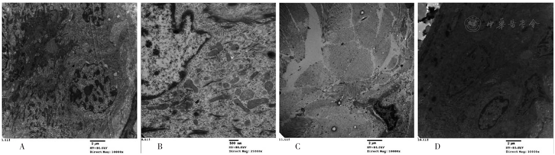
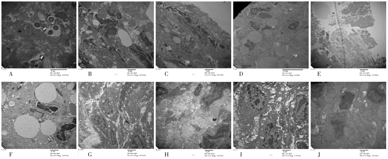
摘要: 背景 压疮又称褥疮、席疮,对应现代医学压力性损伤,是由于局部组织长期受压,继而发生持续缺血、缺氧以及营养不良,导致组织发生溃烂、坏死的外科疾病。艾灸治疗压疮临床效果显著,但相关基础研究较少,其机制尚不清楚。 目的 通过透射电镜观察回旋灸对压力性损伤大鼠创面组织超微结构的影响,明确回旋灸促进创面修复作用,以期为临床艾灸治疗压力性损伤提供理论依据和参考方案。 方法 于2020年12月至2021年11月,选取120只健康雌性SD成年大鼠,其中85只使用自制造模装置通过缺血-再灌注损伤方式建立2~3期压力性损伤大鼠模型,将模型制备成功的70只雌性SD大鼠随机分为回旋灸组(n=35)与模型对照组(n=35),另选取35只健康雌性SD大鼠作为空白对照组,每组根据干预时间分为1、3、5、7、10 d 5个亚组,每亚组7只。回旋灸组碘伏处理后给予回旋灸治疗(1次/d,15 min/次),模型对照组和空白对照组仅进行碘伏常规处理。分别于干预的1、3、5、7、10 d对各亚组大鼠取样,采用透射电镜观察压力性损伤皮肤组织的超微结构。 结果 电镜观察结果显示,与空白对照组比较,大鼠造模后表皮脱落或萎缩,原有正常皮肤结构发生改变。模型对照组不同时间点呈现了自身修复中的基本病理状态变化:5 d亚组显示为急性炎症的高度浸润阶段,存在大量的炎性细胞高度浸润,以中性粒细胞为主,此时期的中性粒细胞为新鲜状态;10 d亚组镜下可见少部分表皮结构完整,大部分未见全层表皮结构,仅存在基底层及棘层,缺失颗粒层、透明层及角质层,基底细胞与棘细胞的线粒体仍呈现肿胀状态,炎性细胞浸润以凋亡的淋巴细胞与吞噬细胞为主。回旋灸组随着艾灸干预刺激的累积,表皮结构逐渐修复:5 d亚组可见表皮结构,部分完整,部分仅可见基底层及棘层;7 d亚组可见表皮的完整全层结构,基底层、棘层、颗粒层、透明层、角质层均清晰可见;5~10 d亚组基底细胞及棘细胞的线粒体的结构、数量、形态由损伤后的肿胀、数量较少、不丰富、结构不清晰逐渐向修复后的不肿胀、数量较多、丰富、结构清晰的方向发展转变。回旋灸组炎性细胞镜下所示:1 d亚组为大量中性粒细胞浸润;3 d亚组为中等量的中性粒细胞,其炎性细胞数量相比1 d亚组减少,部分炎性细胞处于凋亡状态,核固缩明显;5 d亚组为不新鲜、陈旧性中性粒细胞;7 d亚组为吞噬细胞及淋巴细胞;10 d亚组为淋巴细胞。 结论 (1)回旋灸对压力性损伤大鼠创面组织的表皮修复发挥明显促进作用。(2)回旋灸从整体上提前急性炎症浸润的高峰,在整体进程上缩短创面修复所需时间,有效促进压力性损伤创面的愈合。

关键词: 压力性损伤, 压疮, 回旋灸, 艾灸, 超微结构, 血管新生, 创面修复, 透射电镜观察

Abstract:

Background

Pressure ulcers, also called decubitus ulcers and bedsores, correspond to pressure injuries in modern medicine, which is a surgical disease manifested by tissue ulceration and necrosis due to continuous ischemia, hypoxia and malnutrition associated with long-term pressure. Moxibustion has significant clinical effect on pressure injury, but there are few relevant fundamental researches and its mechanism is still unclear.

Objective

To observe the effect of circling moxibustion on the ultrastructure of wound tissue in rats with pressure injury using transmission electron microscope, clarify the role of circling moxibustion in promoting wound repair, and provide a theoretical basis and reference plan for clinical moxibustion treatment of pressure injuries.

Methods

A total of 120 healthy female SD adult rats were selected from December 2020 to November 2021, and 85 of which were selected to establish the rat model of stage 2 or 3 pressure injury through ischemia-reperfusion injury with self-made modeling device. The 70 female SD rats with successful model preparation were randomly divided into circling moxibustion group (n=35) and model control group (n=35). Another 35 healthy female SD rats were selected as the blank control group. Each group was divided into five subgroups including the 1st, 3rd, 5th, 7th and 10th day according to the length of intervention time, with 7 rats in each subgroup. The circling moxibustion group received circling moxibustion treatment (once /d for 15 min each time) after routine iodophor intervention, while the model control group and blank control group only received routine iodophor intervention. On the 1st, 3rd, 5th, 7th and 10th day of the intervention, the rats in each subgroup were sampled and transmission electron microscope was used to observe the ultrastructure of skin tissue with pressure injury.

Results

The results of transmission electron microscope observation showed that the epidermis of rats was exfoliated or atrophied after modeling and the original normal skin structure was changed compared with blank control group. The model control group showed changes in the fundamental pathological state of self-repair at different time-points: 5 d subgroup showed a high infiltration stage of acute inflammation with a large number of inflammatory cells highly infiltrated which mainly belonged to neutrophils in fresh state. 10 d subgroup showed that only a small part of the epidermal structure was intact and most of the epidermal structure could not be seen, which only presented stratum basale and stratum spinosum without stratum granulosum, stratum lucidum and stratum corneum. The mitochondria of basal cells and prickle cells were still swollen, and the inflammatory cells were mainly infiltrated by apoptotic lymphocytes and phagocytes. With the accumulation of moxibustion intervention stimulation, the epidermis structure in the circling moxibustion group gradually repaired: 5 d subgroup showed that the epidermal structure was partially intact, only stratum basale and stratum spinosum could be seen partially. The complete full-thickness structures of the epidermis including stratum basale, stratum spinosum, stratum granulosum, stratum lucidum and stratum corneum existed in the 7 d subgroup. 5-10 d subgroups showed that the structure, quantity and morphology of mitochondria of basal cells and spinous cells gradually changed from the state of swelling, less quantity, less abundance and unclear structure after injury to the direction of no swelling, more quantity, abundance and clear structure after repair. The results of circling moxibustion group under the microscope of inflammatory cells showed that there were substantial neutrophils infiltrations in the 1d subgroup, which decreased to the medium amount in the 3 d subgroup with fewer inflammatory cells. Several inflammatory cells were in the state of apoptosis with obvious karyopyknosis. There were stale and old neutrophils in the 5 d subgroup, phagocytes and lymphocytes in the 7 d subgroup and lymphocytes in the 10 d subgroup.

Conclusion

(1) Circling moxibustion can play a significant role in promoting epidermal repair of the wound tissue in rats with pressure injury. (2) Circling moxibustion can make the peak of acute inflammatory infiltration in advance and shorten the time required for wound repair in the overall process as a whole, indicating that circling moxibustion can effectively promote the wound healing in rats with pressure injury.

Key words: Pressure injury, Pressure ulcer, Circling moxibustion, Moxibustion, Ultrastructure, Angiogenesis, Wound repair, Transmission electron microscope observation