中国全科医学 ›› 2022, Vol. 25 ›› Issue (24): 3065-3069.DOI: 10.12114/j.issn.1007-9572.2022.0185

• 典型病例 • 上一篇    下一篇

中性脂肪沉积症伴肌病患者的外周血细胞形态特点并文献复习

邢莹, 普程伟, 尚柯, 屈晨雪*()   

  1. 100034 北京市,北京大学第一医院检验科
  • 收稿日期:2022-01-11 修回日期:2022-03-26 出版日期:2022-08-20 发布日期:2022-06-02
  • 通讯作者: 屈晨雪
  • 邢莹,普程伟,尚柯,等.中性脂肪沉积症伴肌病患者的外周血细胞形态特点并文献复习[J].中国全科医学,2022,25(24):3065-3069.[www.chinagp.net]
    作者贡献:邢莹设计研究方案、负责论文起草;普程伟、尚柯负责进行实验及图片采集;屈晨雪负责最终版本修订,对论文负责。

Morphological Characteristics of Peripheral Blood Cells in Patients with Neutral Lipid Storage Disease with Myopathy and Literature Review

Ying XING, Chengwei PU, Ke SHANG, Chenxue QU*()   

  1. Laboratory Department, Peking University First Hospital, Beijing100034, China
  • Received:2022-01-11 Revised:2022-03-26 Published:2022-08-20 Online:2022-06-02
  • Contact: Chenxue QU
  • About author:
    XING Y, PU C W, SHANG K, et al. Morphological characteristics of peripheral blood cells in patients with neutral lipid storage disease with myopathy and literature review[J]. Chinese General Practice, 2022, 25 (24) : 3065-3069.

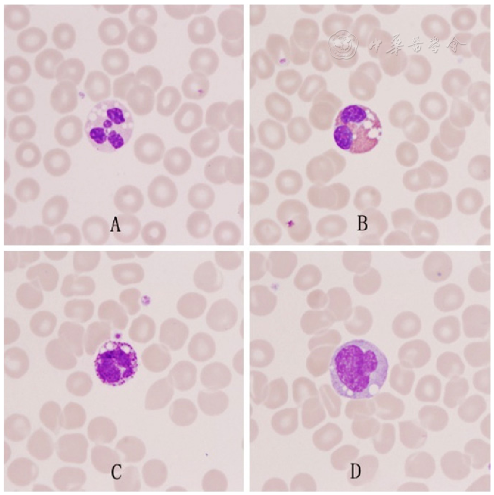
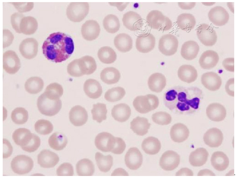
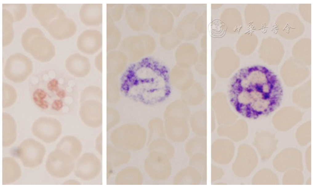
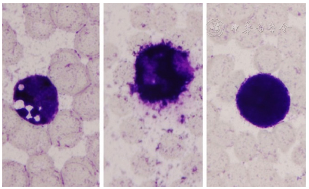
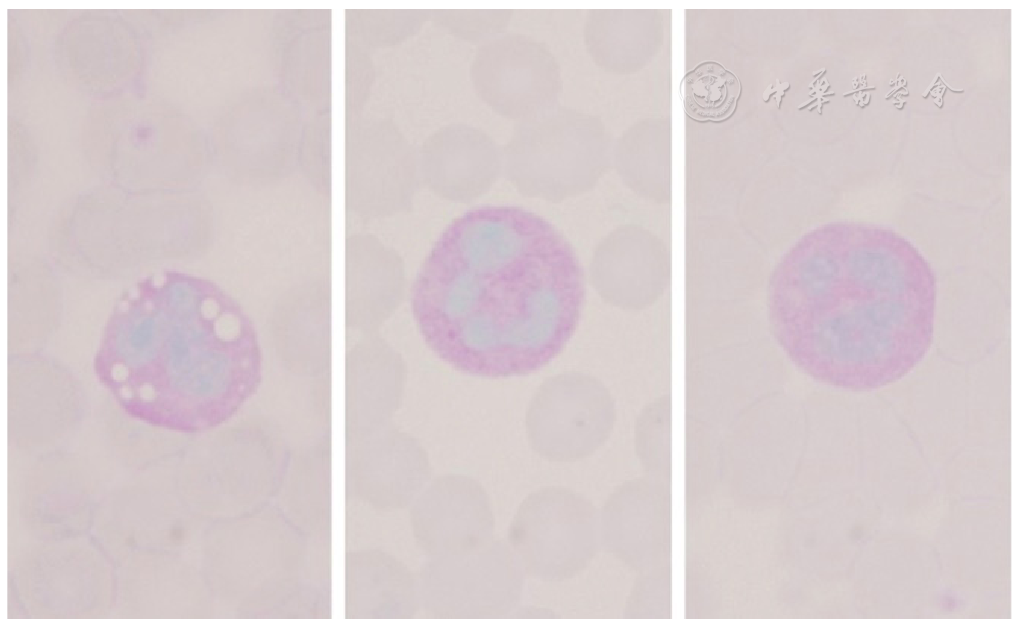
摘要: 背景 中性脂肪沉积症伴肌病(NLSDM)起病隐匿、症状缺乏特异性,由于患者临床表现不同,可能就诊于神经科、心血管疾病科等多个科室,容易导致漏诊和误诊。其明确诊断有赖于基因检测,但多数患者由于不能及时明确诊断而延误治疗。此类患者外周血白细胞具有典型的形态学特征,可提示临床医生及时完善基因检测以明确诊断。 目的 探讨NLSDM患者的外周血细胞形态特点。 方法 选取2021年6—8月北京大学第一医院收治的3例NLSDM患者为研究对象,收集患者外周血并制备血涂片,分别采用瑞-姬氏染色、中性粒细胞碱性磷酸酶染色(NAP)、髓过氧化物酶染色(MPO)和糖原染色(PAS)观察血细胞形态特点。 结果 瑞-姬氏染色:中性粒细胞、嗜酸粒细胞、嗜碱粒细胞及单核细胞胞质中均可见数个大小不一的圆形空泡。NAP染色:NLSDM患者的中性粒细胞胞质中呈阴性反应,与正常人和感染患者差异明显,感染患者阳性率和积分值均明显增高;MPO染色:NLSDM患者的中性粒细胞胞质中呈阳性反应,与正常人的中性粒细胞阳性程度接近,但弱于感染患者(阳性颗粒粗大并覆盖在细胞核上,导致细胞核结构不清);PAS:NLSDM患者、正常人及感染患者的中性粒细胞阳性反应程度均未见明显差异。 结论 NLSDM患者外周血多种白细胞胞质中均可见大小不等的空泡,而中性粒细胞空泡与感染时的中毒性改变不同,通过NAP可对其初步鉴别,这种形态学异常是NLSDM的特征性改变。

关键词: 脂质贮积病, 中性脂肪沉积症伴肌病, 中性粒细胞中毒性改变, 中性粒细胞碱性磷酸酶染色

Abstract:

Background

Neutral lipid storage disease with myopathy (NLSDM) has obscure onset and nonspecific clinical symptoms. Due to different clinical manifestations, the patients with NLSDM may be seen in neurology, cardiovascular disease and other departments, which may easily lead to missed diagnosis and misdiagnosis. The definitive diagnosis depends on genetic detection, but most patients delay treatment because they cannot be diagnosed in time. The peripheral blood leukocytes of such patients have typical morphological characteristics, which can prompt clinicians to improve genetic testing in time to confirm the diagnosis.

Objective

To investigate the morphological characteristics of peripheral blood cells in NLSDM patients.

Methods

3 NLSDM patients admitted to Peking University First Hospital from June to August 2021 were selected as the subjects, the peripheral blood of the patients was collected to prepare blood smears, and the morphological characteristics of blood cells were observed by Wright-Giemsa staining, neutrophil alkaline phosphatase (NAP) staining, myeloperoxidase (MPO) staining and periodic acid-schiff reaction (PAS) .

Results

Wright-Giemsa staining: several circular vacuoles of different sizes can be seen in the cytoplasm of neutrophils, eosinophils, basophils, and monocytes; NAP: the neutrophil cytoplasm of NLSDM patients was negative, which was significantly different from normal and infected patients, the positive rate and integral value of infected patients were significantly increased; MPO: patients with NLSDM were positive which were similar to healthy individual but weaker than infected patients (the large positive particles covered the nucleus, resulting in unclear nuclear structure) . PAS: there was no significant difference in the positive degree of NLSDM patients, healthy individual and infected patients.

Conclusion

Vacuoles of different sizes can be seen in the cytoplasm of various leukocytes in the peripheral blood of NLSDM patients, while neutrophil vacuoles are different from the toxic changes during infection, which can be preliminarily identified by NAP staining. Such morphological abnormalities are characteristic changes of NLSDM.

Key words: Lipidoses, Neutral lipid storage disease with myopathy, Toxic changes of neutrophils, Neutrophil alkaline phosphatase staining