
Chinese General Practice ›› 2025, Vol. 28 ›› Issue (11): 1336-1341.DOI: 10.12114/j.issn.1007-9572.2024.0408
Special Issue: 消化系统疾病最新文章合辑
• Original Research • Previous Articles Next Articles
Received:2024-08-03
Revised:2024-11-10
Published:2025-04-15
Online:2025-02-06
Contact:
GU Tianwei
通讯作者:
顾天伟
作者简介:徐浩负责临床数据收集、整理、分析,并撰写论文初稿;方达负责绘制图表并协助统计分析;周卫红负责体检人群的检测;毕艳完善论文的审校;顾天伟提出研究思路,设计研究方案,完善论文最终内容及审校,并对论文负责。
基金资助:CLC Number:
Add to citation manager EndNote|Ris|BibTeX
URL: https://www.chinagp.net/EN/10.12114/j.issn.1007-9572.2024.0408
| 项目 | 未发生脂肪肝组(n=14 563) | 发生脂肪肝组(n=2 523) | t(χ2)值 | P值 |
|---|---|---|---|---|
| 性别(男/女) | 7 624/6 939 | 1 837/686 | 364.226a | <0.001 |
| 年龄(岁) | 43.9±13.2 | 46.6±13.4 | -9.384 | <0.001 |
| BMI(kg/m2) | 22.9±2.5 | 25.0±2.6 | -37.979 | <0.001 |
| WC(cm) | 78.30±8.82 | 85.50±8.01 | -41.081 | <0.001 |
| SBP(mmHg) | 124±17 | 129±17 | -14.368 | <0.001 |
| DBP(mmHg) | 76±11 | 80±14 | -14.717 | <0.001 |
| FBG(mmol/L) | 4.94±0.88 | 5.15±1.02 | -10.049 | <0.001 |
| HbA1c(%) | 5.56±0.62 | 5.70±0.71 | -6.064 | <0.001 |
| ALT(U/L) | 19.53±17.40 | 23.84±14.37 | -13.476 | <0.001 |
| AST(U/L) | 19.92±10.62 | 20.63±6.79 | -3.243 | <0.001 |
| TG(mmol/L) | 1.02±0.64 | 1.44±0.87 | -23.101 | <0.001 |
| TC(mmol/L) | 4.61±0.87 | 4.71±0.91 | -4.986 | <0.001 |
| HDL-C(mmol/L) | 1.46±0.37 | 1.24±0.30 | 32.485 | <0.001 |
| LDL-C(mmol/L) | 2.68±0.73 | 2.84±0.75 | -10.105 | <0.001 |
| VAI | 1.17±1.15 | 1.81±1.51 | -20.554 | <0.001 |
| CVAI | 57.30±40.91 | 92.12±35.04 | -44.892 | <0.001 |
Table 1 Comparison of baseline clinical data between the fatty liver group and non-fatty liver group
| 项目 | 未发生脂肪肝组(n=14 563) | 发生脂肪肝组(n=2 523) | t(χ2)值 | P值 |
|---|---|---|---|---|
| 性别(男/女) | 7 624/6 939 | 1 837/686 | 364.226a | <0.001 |
| 年龄(岁) | 43.9±13.2 | 46.6±13.4 | -9.384 | <0.001 |
| BMI(kg/m2) | 22.9±2.5 | 25.0±2.6 | -37.979 | <0.001 |
| WC(cm) | 78.30±8.82 | 85.50±8.01 | -41.081 | <0.001 |
| SBP(mmHg) | 124±17 | 129±17 | -14.368 | <0.001 |
| DBP(mmHg) | 76±11 | 80±14 | -14.717 | <0.001 |
| FBG(mmol/L) | 4.94±0.88 | 5.15±1.02 | -10.049 | <0.001 |
| HbA1c(%) | 5.56±0.62 | 5.70±0.71 | -6.064 | <0.001 |
| ALT(U/L) | 19.53±17.40 | 23.84±14.37 | -13.476 | <0.001 |
| AST(U/L) | 19.92±10.62 | 20.63±6.79 | -3.243 | <0.001 |
| TG(mmol/L) | 1.02±0.64 | 1.44±0.87 | -23.101 | <0.001 |
| TC(mmol/L) | 4.61±0.87 | 4.71±0.91 | -4.986 | <0.001 |
| HDL-C(mmol/L) | 1.46±0.37 | 1.24±0.30 | 32.485 | <0.001 |
| LDL-C(mmol/L) | 2.68±0.73 | 2.84±0.75 | -10.105 | <0.001 |
| VAI | 1.17±1.15 | 1.81±1.51 | -20.554 | <0.001 |
| CVAI | 57.30±40.91 | 92.12±35.04 | -44.892 | <0.001 |
| 变量 | 模型1 | 模型2 | 模型3 | |||
|---|---|---|---|---|---|---|
| HR(95%CI) | P值 | HR(95%CI) | P值 | HR(95%CI) | P值 | |
| VAI | ||||||
| Q1 | 1.000 | 1.000 | 1.000 | |||
| Q2 | 1.829(1.545~2.164) | <0.001 | 1.499(1.266~1.776) | <0.001 | 1.409(1.180~1.681) | <0.001 |
| Q3 | 3.380(2.897~3.944) | <0.001 | 2.312(1.975~2.706) | <0.001 | 2.051(1.715~2.454) | <0.001 |
| Q4 | 5.734(4.949~6.643) | <0.001 | 3.204(2.744~3.742) | <0.001 | 2.579(2.088~3.186) | <0.001 |
| CVAI | ||||||
| Q1 | 1.000 | 1.000 | 1.000 | |||
| Q2 | 3.548(2.877~4.376) | <0.001 | 3.167(2.533~3.961) | <0.001 | 2.431(1.939~3.048) | <0.001 |
| Q3 | 6.689(5.484~8.158) | <0.001 | 5.300(4.163~6.747) | <0.001 | 3.335(2.599~4.278) | <0.001 |
| Q4 | 11.241(9.263~13.642) | <0.001 | 6.897(5.169~9.202) | <0.001 | 3.375(2.488~4.576) | <0.001 |
Table 2 Cox regression analysis of the correlation of VAI and CVAI with fatty liver development
| 变量 | 模型1 | 模型2 | 模型3 | |||
|---|---|---|---|---|---|---|
| HR(95%CI) | P值 | HR(95%CI) | P值 | HR(95%CI) | P值 | |
| VAI | ||||||
| Q1 | 1.000 | 1.000 | 1.000 | |||
| Q2 | 1.829(1.545~2.164) | <0.001 | 1.499(1.266~1.776) | <0.001 | 1.409(1.180~1.681) | <0.001 |
| Q3 | 3.380(2.897~3.944) | <0.001 | 2.312(1.975~2.706) | <0.001 | 2.051(1.715~2.454) | <0.001 |
| Q4 | 5.734(4.949~6.643) | <0.001 | 3.204(2.744~3.742) | <0.001 | 2.579(2.088~3.186) | <0.001 |
| CVAI | ||||||
| Q1 | 1.000 | 1.000 | 1.000 | |||
| Q2 | 3.548(2.877~4.376) | <0.001 | 3.167(2.533~3.961) | <0.001 | 2.431(1.939~3.048) | <0.001 |
| Q3 | 6.689(5.484~8.158) | <0.001 | 5.300(4.163~6.747) | <0.001 | 3.335(2.599~4.278) | <0.001 |
| Q4 | 11.241(9.263~13.642) | <0.001 | 6.897(5.169~9.202) | <0.001 | 3.375(2.488~4.576) | <0.001 |
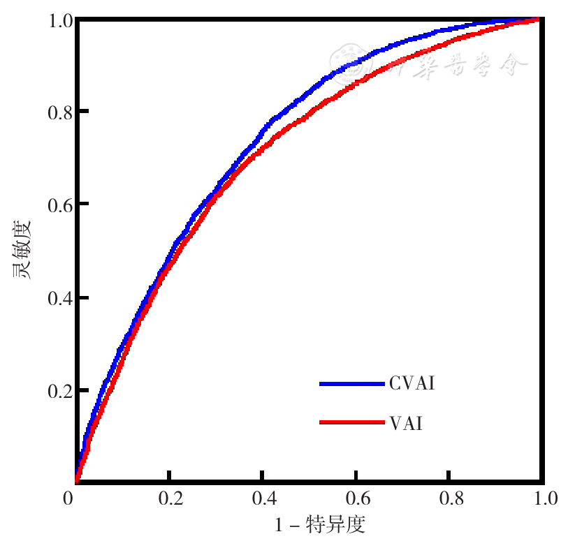
Figure 1 ROC curves of VAI and CVAI in predicting fatty liver
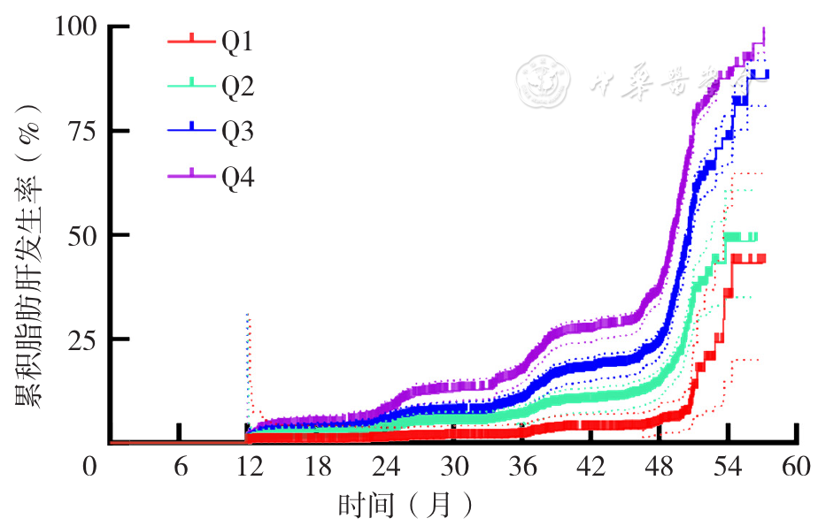
Figure 2 Kaplan-Meier survival curves for subjects divided by the quartiles of CVAI
| 分层 | CVAI-Q1 | CVAI-Q2 | P值 | CVAI-Q3 | P值 | CVAI-Q4 | P值 |
|---|---|---|---|---|---|---|---|
| 性别 | |||||||
| 男 | 1.00 | 1.98(1.22~3.21) | 0.006 | 2.53(1.57~4.07) | <0.001 | 4.12(2.57~6.58) | <0.001 |
| 女 | 1.00 | 1.34(0.88~2.06) | 0.174 | 1.54(0.78~3.07) | 0.215 | 2.54(1.20~5.38) | 0.015 |
| 年龄 | |||||||
| 18~<40岁 | 1.00 | 1.81(1.26~2.60) | 0.001 | 1.85(1.14~3.00) | 0.012 | 2.45(1.35~4.46) | 0.003 |
| ≥40岁 | 1.00 | 2.03(1.07~3.84) | 0.029 | 3.37(1.79~6.31) | <0.001 | 8.63(4.73~15.77) | <0.001 |
| BMI | |||||||
| 正常(18~24 kg/m2) | 1.00 | 1.92(1.40~2.64) | <0.001 | 2.20(1.48~3.27) | <0.001 | 2.73(1.43~5.21) | 0.002 |
| 超重(>24 kg/m2) | 1.00 | 1.42(0.69~2.95) | 0.343 | 2.05(1.02~4.12) | 0.045 | 3.63(1.85~7.14) | <0.001 |
Table 3 A hierarchical analysis of the correlation between CVAI and fatty liver
| 分层 | CVAI-Q1 | CVAI-Q2 | P值 | CVAI-Q3 | P值 | CVAI-Q4 | P值 |
|---|---|---|---|---|---|---|---|
| 性别 | |||||||
| 男 | 1.00 | 1.98(1.22~3.21) | 0.006 | 2.53(1.57~4.07) | <0.001 | 4.12(2.57~6.58) | <0.001 |
| 女 | 1.00 | 1.34(0.88~2.06) | 0.174 | 1.54(0.78~3.07) | 0.215 | 2.54(1.20~5.38) | 0.015 |
| 年龄 | |||||||
| 18~<40岁 | 1.00 | 1.81(1.26~2.60) | 0.001 | 1.85(1.14~3.00) | 0.012 | 2.45(1.35~4.46) | 0.003 |
| ≥40岁 | 1.00 | 2.03(1.07~3.84) | 0.029 | 3.37(1.79~6.31) | <0.001 | 8.63(4.73~15.77) | <0.001 |
| BMI | |||||||
| 正常(18~24 kg/m2) | 1.00 | 1.92(1.40~2.64) | <0.001 | 2.20(1.48~3.27) | <0.001 | 2.73(1.43~5.21) | 0.002 |
| 超重(>24 kg/m2) | 1.00 | 1.42(0.69~2.95) | 0.343 | 2.05(1.02~4.12) | 0.045 | 3.63(1.85~7.14) | <0.001 |
| [1] |
|
| [2] |
|
| [3] |
|
| [4] |
|
| [5] |
|
| [6] |
|
| [7] |
|
| [8] |
|
| [9] |
|
| [10] |
|
| [11] |
聂倩,孟翠巧,刘焕欣,等. 不同肥胖指标及内脏脂肪指数对非酒精性脂肪肝预测价值的比较[J]. 中国老年学杂志,2024,44(6):1321-1325.
|
| [12] |
王洪岩,刘宇鹏,付红梅,等. 内脏脂肪指数和脂质蓄积指数对非超重/肥胖者代谢相关脂肪性肝病的预测价值[J]. 中华健康管理学杂志,2023,17(11):848-853. DOI:10.3760/cma.j.cn115624-20230615-00375.
|
| [13] |
|
| [14] |
|
| [15] |
|
| [16] |
|
| [17] |
|
| [18] |
|
| [19] |
|
| [20] |
|
| [21] |
|
| [22] |
|
| [23] |
|
| [24] |
|
| [25] |
|
| [26] |
|
| [27] |
|
| [28] |
|
| [29] |
王丽娜,高鹏飞,曹帆,等. 不同性别人群非酒精性脂肪性肝病患病现况及影响因素分析[J]. 中国全科医学,2023,26(33):4143-4151.
|
| [30] |
|
| [1] | HAN Congcong, QIU Xinyu, SHAN Chunfang, SONG Ning, CHEN Qingjie, MULADILI· Abudureheman, LI Xiaomei, YANG Yining, ZHAO Qian. Impact of Metabolic Obesity Phenotype on Long-term Prognosis after Percutaneous Coronary Intervention in Patients with Acute Coronary Syndrome [J]. Chinese General Practice, 2026, 29(21): 2950-2958. |
| [2] | AN Yanhong, WANG Shidong, LI Xiaoran, GUO Jiayang, WANG Zhe, SHA Peilin, MENG Yijun, LI Xiaoxuan, SHI Xue, YU Zexing, XIAO Yonghua. Study on Risk Factors and Nomogram Prediction Model for Diabetic Kidney Disease: Based on Contrast-enhanced Ultrasound Technology [J]. Chinese General Practice, 2026, 29(21): 2995-3003. |
| [3] | ZHANG Ke, ZENG Xianchang, ZOLZAYA Enkhzaya, ZHU Yelin, HUANG Yiwen, LIU Zhenxiu, TAO Feng. Effect of Xiere Xingpi Yin on Eating Behaviors and Weight-related Outcomes in Patients with Obesity: a Randomized Controlled Trial [J]. Chinese General Practice, 2026, 29(20): 2759-2765. |
| [4] | GE Dan, WANG Zhi, DING Qun, GUO Tonglan, XU Tongdao. Relationship between Remnant Cholesterol and Non-alcoholic Fatty Liver Disease as well as Progressive Liver Fibrosis in Patients with Type 2 Diabetes Mellitus [J]. Chinese General Practice, 2026, 29(20): 2854-2859. |
| [5] | SONG Zhenzhen, CHEN Yang, WANG Jiwen, LIU Huishan, SHEN Simeng, SUN Ying, CHEN Xing. Study of Maternal Risk Factors Associated with Neonatal Asphyxia in Late Preterm and Term Infants [J]. Chinese General Practice, 2026, 29(20): 2874-2878. |
| [6] | YANG Xiaofeng, LI Xianwen, WU Yanfeng, ZHANG Qian, CHEN Juan, ZHAO Fengjiao. Association of Serum Vascular Endothelial Cadherin Levels with the Risk of Post-stroke Cognitive Impairment in Patients with Acute Ischemic Stroke [J]. Chinese General Practice, 2026, 29(20): 2867-2873. |
| [7] | REYILAI· Maimaiti, ZHOU Yiran, WU Yun, LIU Zhencheng, LU Yaoqin, WU Haiyan. Association between Novel Obesity Indicators and Cardiovascular Disease Risk in Hypertensive Patients [J]. Chinese General Practice, 2026, 29(20): 2836-2845. |
| [8] | WU Zhen, XI Yaqi, HU Linlin. Characteristics of Policy Instrument Selection and Combination for Weight Management in China: a Content Analysis of National Policy Documents (2016-2025) [J]. Chinese General Practice, 2026, 29(20): 2766-2774. |
| [9] | CHEN Xiangyang, HUANG Hongmei, LI Sheyu. Prevention and Management of Adult Obesity: Progress and Trends from Six Keywords in 2025 [J]. Chinese General Practice, 2026, 29(20): 2753-2758. |
| [10] | ZHAO Huili, LU Lina, ZHUO Ya, WANG Xin. Clinical Characteristics and Risk Factors for Recurrence of Cryptogenic Organizing Pneumonia [J]. Chinese General Practice, 2026, 29(19): 2648-2655. |
| [11] | TANG Lijuan, QI Qi, ZHANG Fan, GAO Yifu, CAO Yajing, YUE Fujuan, GAO Jinchai, LIU Xiaoli. Characterisation of the Prevalence of Overweight/Obesity among Residents Aged 18-44 Years in Hebei Province in 2013 and 2020 [J]. Chinese General Practice, 2026, 29(19): 2695-2704. |
| [12] | YUAN Yujuan, TAO Jing, WANG Ying, PENG Hui, YANG Yining. Elevated Neutrophil Percentage to Albumin Ratio is Associated with In-hospital Outcomes in Patients with Acute Myocardial Infarction [J]. Chinese General Practice, 2026, 29(18): 2489-2497. |
| [13] | LI Yachan, YANG Yang, XU Qianting, KE Tingyu. Association between the Chinese Visceral Adiposity Index and Left Ventricular Diastolic Dysfunction in Type 2 Diabetes Mellitus [J]. Chinese General Practice, 2026, 29(18): 2482-2488. |
| [14] | LI Simin, ZHANG Tingting, WANG Kunbo, YANG Jianzhou, PING Weiwei. Effects of Exercise Dosage on Elderly Patients with Sarcopenia: a Meta-analysis [J]. Chinese General Practice, 2026, 29(17): 2400-2409. |
| [15] | HUANG Minshan, CHEN Hang, LI Liya, YANG Taiming, CHEN Lifang, LI Shenchao, REN Li, WANG Hui, LI Mingke, WANG Xianmei, WANG Da, WAN Ying, HE Yule, ZHOU Qingqing, LI Yu, LI Mengwei, LU Lihong, LUO Yifan, MA Lanqing. The Current Status of Awareness and Influencing Factors Analysis of Metabolic-associated Fatty Liver Disease among the General Population in Multiple Regions of Yunnan [J]. Chinese General Practice, 2026, 29(17): 2347-2353. |
| Viewed | ||||||
|
Full text |
|
|||||
|
Abstract |
|
|||||