
Chinese General Practice ›› 2025, Vol. 28 ›› Issue (17): 2149-2155.DOI: 10.12114/j.issn.1007-9572.2024.0160
Special Issue: 乳腺癌最新文章合辑
• Original Research • Previous Articles Next Articles
Received:2024-04-21
Revised:2024-08-24
Published:2025-06-15
Online:2025-04-22
Contact:
LI Jun
通讯作者:
李军
作者简介:作者贡献:
邓雅倩负责文章的构思与设计,撰写论文;曹春莉负责研究的实施与可行性分析;邓雅倩、马金梅进行数据收集;李文肖、徐泽林负责数据整理;徐泽林负责统计学处理;邓雅倩、曹春莉负责结果的分析与解释;邓雅倩、马金梅进行论文的修订;成静负责文章的质量控制及审校;李军对文章整体负责,监督管理。
基金资助:CLC Number:
Add to citation manager EndNote|Ris|BibTeX
URL: https://www.chinagp.net/EN/10.12114/j.issn.1007-9572.2024.0160
| 分类 | 个数 | 年龄[M(P25,P75),岁] | 病理类型[例(%)] | |||||
|---|---|---|---|---|---|---|---|---|
| 浸润性导管癌 | 原位癌 | 浸润性小叶癌 | 黏液癌 | 髓样癌 | 乳头状癌 | |||
| 未转移者 | 105 | 51.0(42.0,60.0) | 76(72.4) | 18(17.1) | 4(3.8) | 3(2.9) | 3(2.9) | 1(0.9) |
| 转移者 | 61 | 52.0(39.0,65.0) | 56(91.8) | 2(3.3) | 2(3.3) | 1(1.6) | 0 | 0 |
| χ2(Z)值 | -0.746a | 10.577 | ||||||
| P值 | 0.456 | 0.600 | ||||||
Table 1 Comparison of age and pathological types between axillary lymph node metastatic breast cancer patients and non-metastatic breast cancer patients
| 分类 | 个数 | 年龄[M(P25,P75),岁] | 病理类型[例(%)] | |||||
|---|---|---|---|---|---|---|---|---|
| 浸润性导管癌 | 原位癌 | 浸润性小叶癌 | 黏液癌 | 髓样癌 | 乳头状癌 | |||
| 未转移者 | 105 | 51.0(42.0,60.0) | 76(72.4) | 18(17.1) | 4(3.8) | 3(2.9) | 3(2.9) | 1(0.9) |
| 转移者 | 61 | 52.0(39.0,65.0) | 56(91.8) | 2(3.3) | 2(3.3) | 1(1.6) | 0 | 0 |
| χ2(Z)值 | -0.746a | 10.577 | ||||||
| P值 | 0.456 | 0.600 | ||||||
| 分类 | 个数 | 肿块最大径[例(%)] | 肿块象限[例(%)] | 形状[例(%)] | ||||||
|---|---|---|---|---|---|---|---|---|---|---|
| ≤2 cm | >2 cm | 外上 | 外下 | 乳晕 | 内上 | 内下 | 规则 | 不规则 | ||
| 未转移者 | 105 | 51(48.6) | 54(51.4) | 49(46.7) | 20(19.0) | 4(3.8) | 27(25.7) | 5(4.8) | 15(14.3) | 90(85.7) |
| 转移者 | 61 | 29(47.5) | 32(52.5) | 26(42.6) | 9(14.8) | 2(2.3) | 17(27.9) | 7(11.5) | 4(6.6) | 57(93.4) |
| 检验统计量值 | 0.016 | 3.050 | 2.274 | |||||||
| P值 | 0.898 | 0.549 | 0.132 | |||||||
| 分类 | 边界[例(%)] | 边缘[例(%)] | 钙化[例(%)] | 血流分级[例(%)] | 纵横比[例(%)] | |||||
| 清晰 | 不清晰 | 光整 | 不光整 | 无 | 有 | 0~Ⅰ级 | Ⅱ~Ⅲ级 | ≤1 | >1 | |
| 未转移者 | 40(38.1) | 65(61.9) | 19(18.1) | 86(81.9) | 65(61.9) | 40(38.1) | 63(60.0) | 42(40.0) | 92(87.6) | 13(12.4) |
| 转移者 | 13(21.3) | 48(78.7) | 4(6.6) | 57(93.4) | 26(42.6) | 35(57.4) | 26(42.6) | 35(57.4) | 45(73.8) | 16(26.2) |
| 检验统计量值 | 5.001 | 4.303 | 5.792 | 4.685 | 5.131 | |||||
| P值 | 0.025 | 0.038 | 0.016 | 0.030 | 0.023 | |||||
| 分类 | 内部回声[例(%)] | 后方回声[例(%)] | 生长方向[例(%)] | SWVmax [M(P25,P75),m/s] | SWVmin [M(P25,P75),m/s] | SWVcentre ( | SWVmean [M(P25,P75),m/s] | |||
| 均匀 | 不均匀 | 未衰减 | 衰减或消失 | 平行 | 不平行 | |||||
| 未转移者 | 7(6.7) | 98(93.3) | 57(54.3) | 48(45.7) | 78(74.3) | 27(25.7) | 5.8(4.6,6.9) | 2.9(1.3,4.5) | 4.1±0.9 | 4.2(3.7,4.7) |
| 转移者 | 5(8.2) | 56(91.8) | 27(44.3) | 34(55.7) | 37(60.7) | 24(39.3) | 7.9(6.0,9.8) | 3.3(2.4,4.2) | 4.7±0.9 | 4.9(4.5,5.3) |
| 检验统计量值 | 0.003 | 1.551 | 3.369 | -9.210a | -1.226a | -4.083b | -8.537a | |||
| P值 | 0.955 | 0.213 | 0.066 | <0.001 | 0.220 | <0.001 | <0.001 | |||
Table 2 Comparison of conventional ultrasound, S-Detect characteristics and VTIQ indexes between axillary lymph node metastatic breast cancer patients and non-metastatic breast cancer patients
| 分类 | 个数 | 肿块最大径[例(%)] | 肿块象限[例(%)] | 形状[例(%)] | ||||||
|---|---|---|---|---|---|---|---|---|---|---|
| ≤2 cm | >2 cm | 外上 | 外下 | 乳晕 | 内上 | 内下 | 规则 | 不规则 | ||
| 未转移者 | 105 | 51(48.6) | 54(51.4) | 49(46.7) | 20(19.0) | 4(3.8) | 27(25.7) | 5(4.8) | 15(14.3) | 90(85.7) |
| 转移者 | 61 | 29(47.5) | 32(52.5) | 26(42.6) | 9(14.8) | 2(2.3) | 17(27.9) | 7(11.5) | 4(6.6) | 57(93.4) |
| 检验统计量值 | 0.016 | 3.050 | 2.274 | |||||||
| P值 | 0.898 | 0.549 | 0.132 | |||||||
| 分类 | 边界[例(%)] | 边缘[例(%)] | 钙化[例(%)] | 血流分级[例(%)] | 纵横比[例(%)] | |||||
| 清晰 | 不清晰 | 光整 | 不光整 | 无 | 有 | 0~Ⅰ级 | Ⅱ~Ⅲ级 | ≤1 | >1 | |
| 未转移者 | 40(38.1) | 65(61.9) | 19(18.1) | 86(81.9) | 65(61.9) | 40(38.1) | 63(60.0) | 42(40.0) | 92(87.6) | 13(12.4) |
| 转移者 | 13(21.3) | 48(78.7) | 4(6.6) | 57(93.4) | 26(42.6) | 35(57.4) | 26(42.6) | 35(57.4) | 45(73.8) | 16(26.2) |
| 检验统计量值 | 5.001 | 4.303 | 5.792 | 4.685 | 5.131 | |||||
| P值 | 0.025 | 0.038 | 0.016 | 0.030 | 0.023 | |||||
| 分类 | 内部回声[例(%)] | 后方回声[例(%)] | 生长方向[例(%)] | SWVmax [M(P25,P75),m/s] | SWVmin [M(P25,P75),m/s] | SWVcentre ( | SWVmean [M(P25,P75),m/s] | |||
| 均匀 | 不均匀 | 未衰减 | 衰减或消失 | 平行 | 不平行 | |||||
| 未转移者 | 7(6.7) | 98(93.3) | 57(54.3) | 48(45.7) | 78(74.3) | 27(25.7) | 5.8(4.6,6.9) | 2.9(1.3,4.5) | 4.1±0.9 | 4.2(3.7,4.7) |
| 转移者 | 5(8.2) | 56(91.8) | 27(44.3) | 34(55.7) | 37(60.7) | 24(39.3) | 7.9(6.0,9.8) | 3.3(2.4,4.2) | 4.7±0.9 | 4.9(4.5,5.3) |
| 检验统计量值 | 0.003 | 1.551 | 3.369 | -9.210a | -1.226a | -4.083b | -8.537a | |||
| P值 | 0.955 | 0.213 | 0.066 | <0.001 | 0.220 | <0.001 | <0.001 | |||

Figure 1 Invasive ductal carcinoma of the breast with axillary lymph node metastasis routine ultrasound,S-Detect examination,VTIQ examination and pathological findings

Figure 2 Invasive ductal carcinoma of the breast without axillary lymph node metastasis routine ultrasound,S-Detect examination,VTIQ examination and pathological findings
| 自变量 | 赋值 |
|---|---|
| 边界 | 清楚=0,不清楚=1 |
| 边缘 | 光整=0,不光整=1 |
| 钙化 | 无=0,有=1 |
| 纵横比 | ≤1=0,>1=1 |
| 血流分级 | 0~Ⅰ级=0,Ⅱ~Ⅲ级=1 |
| SWVmax | 连续变量 |
| SWVcentre | 连续变量 |
| SWVmean | 连续变量 |
Table 3 Assignments for multivariate Logistic regression analysis of influencing factors for axillary lymph node metastasis in breast cancer
| 自变量 | 赋值 |
|---|---|
| 边界 | 清楚=0,不清楚=1 |
| 边缘 | 光整=0,不光整=1 |
| 钙化 | 无=0,有=1 |
| 纵横比 | ≤1=0,>1=1 |
| 血流分级 | 0~Ⅰ级=0,Ⅱ~Ⅲ级=1 |
| SWVmax | 连续变量 |
| SWVcentre | 连续变量 |
| SWVmean | 连续变量 |
| 变量 | B | SE | Wald χ2值 | P值 | OR值(95%CI) |
|---|---|---|---|---|---|
| 边界 | 1.664 | 0.232 | 0.845 | 0.003 | 2.998(1.488~5.085) |
| 边缘 | 1.315 | 0.250 | 5.581 | 0.018 | 3.339(1.473~10.140) |
| 钙化 | 1.757 | 0.947 | 8.470 | 0.004 | 2.759(1.461~8.645) |
| 纵横比 | 1.341 | 0.286 | 6.749 | 0.009 | 8.244(2.271~31.242) |
| 血流分级 | 1.196 | 0.817 | 2.143 | 0.143 | 3.308(0.667~9.913) |
| SWVmax | 0.736 | 0.409 | 0.772 | 0.052 | 1.424(0.275~3.931) |
| SWVcentre | -3.942 | 2.473 | 2.540 | 0.111 | 0.019(0~2.475) |
| SWVmean | 0.710 | 0.458 | 2.885 | 0.005 | 2.173(1.773~4.775) |
Table 4 Multivariate Logistic regression analysis of influencing factors for axillary lymph node metastasis in breast cancer
| 变量 | B | SE | Wald χ2值 | P值 | OR值(95%CI) |
|---|---|---|---|---|---|
| 边界 | 1.664 | 0.232 | 0.845 | 0.003 | 2.998(1.488~5.085) |
| 边缘 | 1.315 | 0.250 | 5.581 | 0.018 | 3.339(1.473~10.140) |
| 钙化 | 1.757 | 0.947 | 8.470 | 0.004 | 2.759(1.461~8.645) |
| 纵横比 | 1.341 | 0.286 | 6.749 | 0.009 | 8.244(2.271~31.242) |
| 血流分级 | 1.196 | 0.817 | 2.143 | 0.143 | 3.308(0.667~9.913) |
| SWVmax | 0.736 | 0.409 | 0.772 | 0.052 | 1.424(0.275~3.931) |
| SWVcentre | -3.942 | 2.473 | 2.540 | 0.111 | 0.019(0~2.475) |
| SWVmean | 0.710 | 0.458 | 2.885 | 0.005 | 2.173(1.773~4.775) |

Figure 3 ROC curves of breast cancer tumors with poorly defined boundaries, uneven edges, aspect ratio >1, calcification, and high SWVmean alone and in combination predicted axillary lymph node metastasis of breast cancer
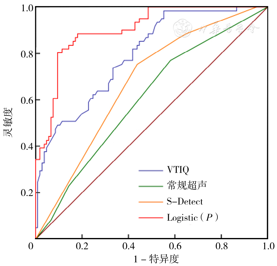
Figure 4 ROC curves of ultrasound,S-Detect and VTIQ alone and in combination predicted axillary lymph node metastasis of breast cancer
| 预测结果 | 未转移者(n=105) | 转移者(n=61) | Kappa值 | P值 | ||
|---|---|---|---|---|---|---|
| 转移 | 未转移 | 转移 | 未转移 | |||
| VTIQ | 14 | 91 | 51 | 10 | 0.693 | <0.005 |
| S-Detect | 18 | 87 | 49 | 12 | 0.619 | <0.005 |
| 常规超声 | 20 | 87 | 47 | 14 | 0.568 | <0.005 |
| Logistic(P) | 13 | 92 | 53 | 8 | 0.732 | <0.005 |
Table 5 Prediction of metastasis by each independent and combine prediction model versus pathological findings
| 预测结果 | 未转移者(n=105) | 转移者(n=61) | Kappa值 | P值 | ||
|---|---|---|---|---|---|---|
| 转移 | 未转移 | 转移 | 未转移 | |||
| VTIQ | 14 | 91 | 51 | 10 | 0.693 | <0.005 |
| S-Detect | 18 | 87 | 49 | 12 | 0.619 | <0.005 |
| 常规超声 | 20 | 87 | 47 | 14 | 0.568 | <0.005 |
| Logistic(P) | 13 | 92 | 53 | 8 | 0.732 | <0.005 |
| [1] |
|
| [2] |
|
| [3] |
|
| [4] |
|
| [5] |
|
| [6] |
|
| [7] |
张冬雪,李卓琳,李振辉,等. 基于DCE-MRI及临床病理特征的模型预测乳腺癌前哨淋巴结状态[J]. 放射学实践,2022,37(9):1104-1108. DOI:10.13609/j.cnki.1000-0313.2022.09.009.
|
| [8] |
桑田. 超声联合免疫组化预测乳腺癌腋窝淋巴结转移的价值[D]. 石河子:石河子大学,2022.
|
| [9] |
|
| [10] |
汪媛媛. 乳腺癌腋窝淋巴结转移的影像及影像组学研究进展[J]. 放射学实践,2023,38(5):662-666. DOI:10.13609/j.cnki.1000-0313.2023.05.024.
|
| [11] |
黄华芳,刘珍. 超声弹性成像联合灰阶超声在乳腺癌腋窝淋巴结转移中的诊断价值[J]. 影像研究与医学应用,2023,7(9):13-15. DOI:10.3969/j.issn.2096-3807.2023.09.005.
|
| [12] |
|
| [13] |
|
| [14] |
向永涛,甘兵,赵亮. 乳腺癌原发灶超声特征与腋窝淋巴结转移的关系[J]. 中国免疫学杂志,2019,35(10):1251-1254. DOI:10.3969/j.issn.1000-484X.2019.10.021.
|
| [15] |
|
| [16] |
|
| [17] |
|
| [18] |
|
| [19] |
姚东. 乳腺癌发生腋窝淋巴结转移与多项超声特征的关系[D]. 衡阳:南华大学,2019.
|
| [20] |
|
| [21] |
|
| [22] |
|
| [23] |
|
| [24] |
吴楠楠,温卫琴,王文娟,等. 剪切波弹性成像联合S-Detect智能辅助诊断技术在甲状腺微小乳头状癌淋巴结转移中的诊断价值[J]. 中国临床实用医学,2021,12(4):43-47. DOI:10.3760/cma.j.cn115570-20210331.00937.
|
| [25] |
杨灵敏.分析超声诊断乳腺癌腋窝淋巴结转移的影像学表现及其临床效果[J]. 中国医药指南,2023,21(9):106-108. DOI:10.15912/j.cnki.gocm.2023.09.012.
|
| [26] |
|
| [27] |
|
| [28] |
|
| [29] |
|
| [30] |
兰梦. 声触诊组织成像定量技术与常规超声在乳腺癌腋窝淋巴结良恶性诊断中的价值[D]. 济南:山东第一医科大学,2019.
|
| Viewed | ||||||
|
Full text |
|
|||||
|
Abstract |
|
|||||