
Chinese General Practice ›› 2022, Vol. 25 ›› Issue (30): 3761-3767.DOI: 10.12114/j.issn.1007-9572.2022.0307
• Article • Previous Articles Next Articles
Received:2022-03-15
Revised:2022-05-19
Published:2022-10-20
Online:2022-06-09
Contact:
Jing MAO
About author:
通讯作者:
毛静
作者简介:基金资助:
Add to citation manager EndNote|Ris|BibTeX
URL: https://www.chinagp.net/EN/10.12114/j.issn.1007-9572.2022.0307
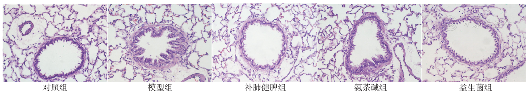
Figure 1 Pathological status in lung tissue of rats in five groups
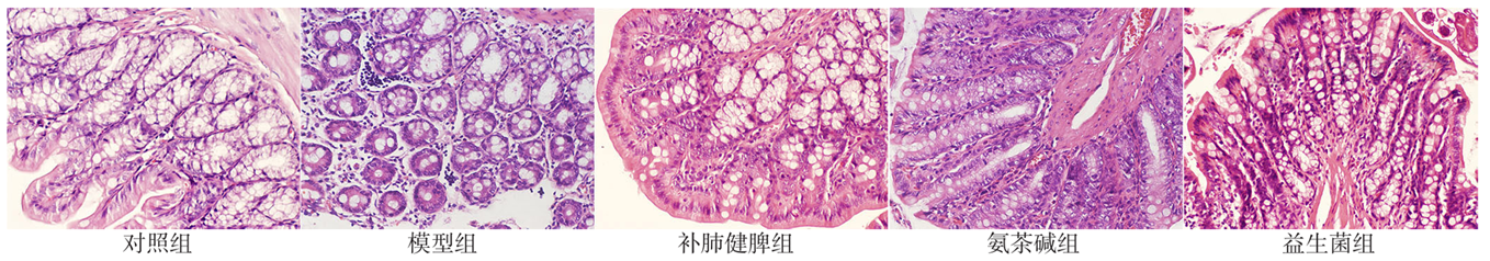
Figure 2 Pathological status in colon tissue of rats in five groups
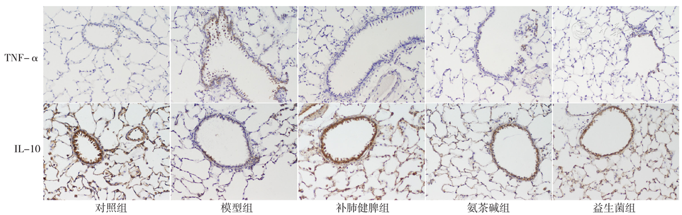
Figure 3 Expression of TNF-α and IL-10 in lung tissue of rats in five groups
| 组别 | 只数 | TNF-α | IL-10 |
|---|---|---|---|
| 对照组 | 6 | 7.39±2.43 | 71.60±18.61 |
| 模型组 | 6 | 47.09±8.22a | 34.78±11.90a |
| 补肺健脾组 | 6 | 7.90±2.38b | 70.72±6.07b |
| 氨茶碱组 | 6 | 14.14±3.09b | 58.98±18.12b |
| 益生菌组 | 6 | 19.17±4.98abc | 45.01±8.38ac |
| F值 | 70.640 | 8.444 | |
| P值 | <0.001 | <0.001 |
Table 1 Expression levels of TNF-α and IL-10 in lung tissue of rats in five groups
| 组别 | 只数 | TNF-α | IL-10 |
|---|---|---|---|
| 对照组 | 6 | 7.39±2.43 | 71.60±18.61 |
| 模型组 | 6 | 47.09±8.22a | 34.78±11.90a |
| 补肺健脾组 | 6 | 7.90±2.38b | 70.72±6.07b |
| 氨茶碱组 | 6 | 14.14±3.09b | 58.98±18.12b |
| 益生菌组 | 6 | 19.17±4.98abc | 45.01±8.38ac |
| F值 | 70.640 | 8.444 | |
| P值 | <0.001 | <0.001 |
| 组别 | 只数 | SIgA |
|---|---|---|
| 对照组 | 6 | 29.00±11.68 |
| 模型组 | 6 | 1.32±0.88a |
| 补肺健脾组 | 6 | 11.34±3.16ab |
| 氨茶碱组 | 6 | 1.59±0.99ac |
| 益生菌组 | 6 | 5.36±1.60a |
| F值 | 26.370 | |
| P值 | <0.001 |
Table 2 Expression levels of SIgA in BALF of rats in five groups
| 组别 | 只数 | SIgA |
|---|---|---|
| 对照组 | 6 | 29.00±11.68 |
| 模型组 | 6 | 1.32±0.88a |
| 补肺健脾组 | 6 | 11.34±3.16ab |
| 氨茶碱组 | 6 | 1.59±0.99ac |
| 益生菌组 | 6 | 5.36±1.60a |
| F值 | 26.370 | |
| P值 | <0.001 |
| 组别 | 只数 | SP | VIP |
|---|---|---|---|
| 对照组 | 6 | 1 361.69±45.07 | 209.81±47.06 |
| 模型组 | 6 | 1 889.38±58.76a | 101.81±63.92a |
| 补肺健脾组 | 6 | 1 469.96±46.74ab | 191.44±40.75b |
| 氨茶碱组 | 6 | 1 842.52±52.37ac | 172.20±39.61 |
| 益生菌组 | 6 | 1 640.42±63.66abcd | 145.26±44.11 |
| F值 | 108.700 | 4.665 | |
| P值 | <0.001 | 0.006 |
Table 3 Expression levels of SP and VIP in lung tissue of rats in five groups
| 组别 | 只数 | SP | VIP |
|---|---|---|---|
| 对照组 | 6 | 1 361.69±45.07 | 209.81±47.06 |
| 模型组 | 6 | 1 889.38±58.76a | 101.81±63.92a |
| 补肺健脾组 | 6 | 1 469.96±46.74ab | 191.44±40.75b |
| 氨茶碱组 | 6 | 1 842.52±52.37ac | 172.20±39.61 |
| 益生菌组 | 6 | 1 640.42±63.66abcd | 145.26±44.11 |
| F值 | 108.700 | 4.665 | |
| P值 | <0.001 | 0.006 |
| 组别 | 只数 | SP | VIP |
|---|---|---|---|
| 对照组 | 6 | 4 786.02±303.29 | 1.42±0.27 |
| 模型组 | 6 | 8 180.00±360.98a | 3.16±0.46a |
| 补肺健脾组 | 6 | 6 585.30±168.88ab | 1.96±0.46b |
| 氨茶碱组 | 6 | 7 587.84±422.13abc | 1.63±0.20b |
| 益生菌组 | 6 | 5 018.25±268.37bcd | 2.38±0.66ab |
| F值 | 136.600 | 13.980 | |
| P值 | <0.001 | <0.001 |
Table 4 Expression levels of SP and VIP in colon tissue of rats in five groups
| 组别 | 只数 | SP | VIP |
|---|---|---|---|
| 对照组 | 6 | 4 786.02±303.29 | 1.42±0.27 |
| 模型组 | 6 | 8 180.00±360.98a | 3.16±0.46a |
| 补肺健脾组 | 6 | 6 585.30±168.88ab | 1.96±0.46b |
| 氨茶碱组 | 6 | 7 587.84±422.13abc | 1.63±0.20b |
| 益生菌组 | 6 | 5 018.25±268.37bcd | 2.38±0.66ab |
| F值 | 136.600 | 13.980 | |
| P值 | <0.001 | <0.001 |
| [1] |
WHO. Global Health Estimates:Life expectancy and leading causes of death and disability[EB/OL]. (2020-11-21)[2022-02-21].
|
| [2] |
赵创艺,袁空军,杨媛,等. 基于ARIMA与NNAR模型的中国慢性阻塞性肺疾病疾病负担预测研究[J]. 中国全科医学,2022,25(16):1942-1949. DOI:10.12114/j.issn.1007-9572.2022.0045.
|
| [3] |
|
| [4] |
谈晓莹,李丹,刘培,等. 基于网络药理学及斑马鱼模型的瓜蒌薤白半夏汤干预慢性阻塞性肺疾病的作用机制研究[J]. 中草药,2021,52(17):5233-5243. DOI:10.7501/j.issn.0253-2670.2021.17.015.
|
| [5] | |
| [6] |
唐诗环,王玥琦,廖强,等. 固本止咳中药对慢性阻塞性肺疾病模型小鼠肺功能及SIgA的影响[J]. 北京中医药,2019,38(9):893-897,945. DOI:10.16025/j.1674-1307.2019.09.014.
|
| [7] |
|
| [8] |
李晓俊,李亚,卞晴晴,等. 中西医结合治疗对慢性阻塞性肺疾病痰热证急性加重-稳定期大鼠免疫因子的影响研究[J]. 中国全科医学,2022,25(2):197-205. DOI:10.12114/j.issn.1007-9572.2021.01.044.
|
| [9] |
|
| [10] |
|
| [11] |
张天宇,张金超,刘妙,等. "通利大肠"对COPD模型大鼠肺组织SP、VIP含量的影响[J]. 世界中医药,2014,9(4):409-411,414. DOI:10.3969/j.issn.1673-7202.2014.04.003.
|
| [12] |
中华中医药学会内科分会肺系病专业委员会. 慢性阻塞性肺疾病中医证候诊断标准(2011版)[J]. 中医杂志,2012,53(2):177-178. DOI:10.13288/j.11-2166/r.2012.02.025.
|
| [13] |
户元元,李亚,李素云,等. 补肺健脾方含药血清对香烟烟雾提取物诱导L6细胞中ROS、MnSOD和CAT表达的影响[J]. 中医杂志,2019,60(7):597-601. DOI:10.13288/j.11-2166/r.2019.07.013.
|
| [14] |
|
| [15] |
|
| [16] |
|
| [17] |
|
| [18] |
|
| [19] |
李晓俊,李亚,李素云. 肠道和肺部微生物群落与慢性阻塞性肺疾病相关性的研究进展[J]. 中国全科医学,2019,22(35):4353-4357. DOI:10.12114/j.issn.1007-9572.2019.00.575.
|
| [20] |
罗珍瑶,梁爱武. SIgA在呼吸道免疫中的作用及中医药干预研究进展[J]. 中西医结合心血管病电子杂志,2019,7(11):72-73. DOI:10.16282/j.cnki.cn11-9336/r.2019.11.054.
|
| [21] |
|
| [22] |
|
| [23] |
中华医学会呼吸病学分会慢性阻塞性肺疾病学组. 慢性阻塞性肺疾病(COPD)中医诊疗指南(2011年修订版)[J]. 中医杂志,2012,1(53):80-84. DOI:10.13288/j.11-2166/r.2012.02.025.
|
| [24] |
|
| [25] |
郑旭锐,杨宇,叶建红,等. 从肺肠病理模型CCK8、CGRP、SP、VIP表达探讨"肺与大肠相表里"[J]. 中成药,2012,34(11):2226-2229. DOI:10.3969/j.issn.1001-1528.2012.11.044.
|
| [26] |
黄子明,侯丽,李蕊白,等. 脾虚证和血管活性肠肽相关性研究进展[J]. 现代中西医结合杂志,2021,30(28):3181-3184. DOI:10.3969/j.issn.1008-8849.2021.28.024.
|
| Viewed | ||||||
|
Full text |
|
|||||
|
Abstract |
|
|||||