中国全科医学 ›› 2023, Vol. 26 ›› Issue (11): 1355-1360.DOI: 10.12114/j.issn.1007-9572.2022.0594

• 论著 • 上一篇    下一篇

mNGS技术和血清G试验在判断耶氏肺孢子菌感染与定植中的价值及二者相关性研究

张彩霞, 刘新年, 杜川, 王新卫*()   

  1. 430033 湖北省武汉市,江汉大学附属湖北省第三人民医院呼吸内科
  • 收稿日期:2022-09-25 修回日期:2022-12-08 出版日期:2023-04-15 发布日期:2022-12-30
  • 通讯作者: 王新卫
  • 张彩霞,刘新年,杜川,等. mNGS技术和血清G试验在判断耶氏肺孢子菌感染与定植中的价值及二者相关性研究[J].中国全科医学,2023,26(11):1355-1360.[www.chinagp.net]

    作者贡献:张彩霞进行文章的构思与设计,文章的可行性分析,文献及资料收集、整理,撰写论文;刘新年进行论文的修订;杜川协助采集数据、分析数据;王新卫负责文章的质量控制及审校,技术支持,对文章整体负责。

mNGS and Serum G Test in Distinguishing between Pneumocystis Jirovecii Colonization and Infection: Value Comparison and Correlation Analysis

ZHANG Caixia, LIU Xinnian, DU Chuan, WANG Xinwei*()   

  1. Department of Respiratory Medicine, Hubei No. 3 People's Hospital of Jianghan University, Wuhan 430033, China
  • Received:2022-09-25 Revised:2022-12-08 Published:2023-04-15 Online:2022-12-30
  • Contact: WANG Xinwei
  • About author:
    ZHANG C X, LIU X N, DU C, et al. mNGS and serum G test in distinguishing between pneumocystis jirovecii colonization and infection: value comparison and correlation analysis [J]. Chinese General Practice, 2023, 26 (11): 1355-1360.

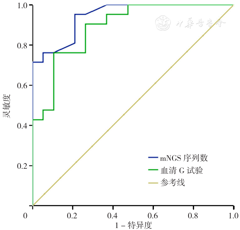
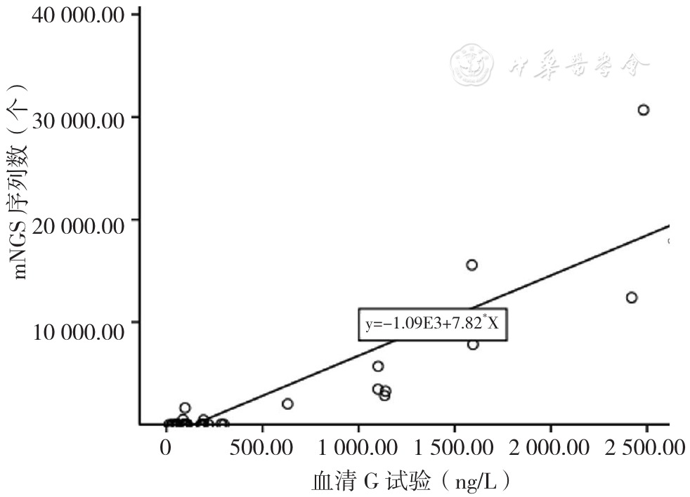
摘要: 背景 耶氏肺孢子菌(PJ)既可定植于肺部,亦可引起肺部感染。为避免临床过度治疗及延误治疗,识别定植与感染很重要。多项研究显示宏基因组二代测序(mNGS)技术及血清1,3-β-D葡聚糖定量检测(G试验)可辅助诊断耶氏肺孢子菌肺炎(PJP),但二者有无鉴别PJ定植和感染的截断值呢? 目的 探讨肺泡灌洗液mNGS技术、血清G试验在判断PJ感染与定植中的价值及二者之间相关性。 方法 收集2018年9月至2022年5月在江汉大学附属湖北省第三人民医院住院治疗,且行肺泡灌洗液mNGS技术检出PJ序列数的40例肺部感染患者病例资料进行回顾性分析。根据mNGS检出PJ是否抗PJ治疗分为PJ感染组21例,PJ定植组19例。收集患者一般资料包括性别、年龄、体质指数(BMI)、吸烟史、基础疾病及用药史〔慢性肾脏疾病、血液系统疾病、自身免疫性疾病、恶性肿瘤、人类免疫缺陷病毒(HIV)感染、实体器官移植、慢性肺部疾病、糖皮质激素/免疫抑制剂使用情况〕,临床症状(发热、咳嗽、咳痰、呼吸困难、胸痛、咯血),影像学特点(磨玻璃渗出影、间质改变、实变、结节、胸腔积液、囊状),实验室指标〔白细胞计数、中性粒细胞计数、淋巴细胞计数、降钙素原(PCT)、C反应蛋白(CRP)、乳酸脱氢酶(LDH)、动脉血氧分压/吸入氧浓度百分比(PaO2/FiO2)、CD4+T淋巴细胞计数〕,绘制mNGS序列数、血清G试验判断PJ感染与定植的受试者工作特征曲线(ROC曲线),分析二者的相关性。 结果 PJ感染组较PJ定植组糖皮质激素/免疫抑制剂使用率高(P<0.05);PJ感染组磨玻璃渗出影、间质改变比例高于PJ定植组(P<0.05);PJ感染组CD4+T淋巴细胞计数低于PJ定植组,而mNGS序列数、血清G试验高于PJ定植组(P<0.05);ROC曲线示mNGS序列数与血清G试验鉴别PJ感染与定植的最佳截断值分别为24个、106.7 ng/L,ROC曲线下面积(AUC)分别为0.95、0.89,灵敏度分别为95.2%、76.2%,特异度分别为78.9%、89.5%。Spearman秩相关结果示mNGS序列数与血清G试验水平呈正相关(rs=0.769、P<0.001)。 结论 PJ感染患者糖皮质激素/免疫抑制剂使用率高,影像学表现为典型磨玻璃渗出影及间质改变,外周血CD4+T淋巴细胞计数下降(尤其<200个/μl),血清G试验及mNGS检出PJ序列数明显升高,分别≥106.7 ng/L、≥24个时,有助于PJ感染的诊断。

关键词: 肺炎,肺囊虫性, 耶氏肺孢子菌, 肺泡灌洗液mNGS技术, 血清G试验, 感染, 定植, 价值, 相关性

Abstract:

Background

Pneumocystis jirovecii (PJ) can colonize the lungs, and cause pulmonary infections. It is essential to distinguish between PJ colonization and infection in the lung to avoid clinical overtreatment and delayed treatment. Many studies have shown that both the mNGS technique and serum G test can be used to aid the diagnosis of PJ pneumonia, but it is still unclear whether they have the appropriate cut-off value to distinguish PJ colonization from PJ infection.

Objective

To investigate the values of mNGS of alveolar lavage fluid and serum G test in distinguishing between infection and colonization of PJ and to assess the correlation between them.

Methods

A retrospective study design was used. Forty inpatients with pulmonary infections were recruited from Hubei No.3 People's Hospital of Jianghan University from September 2018 to May 2022, and divided into PJ infection group (n=21) and colonization group (n=19), according to the prevalence of anti-PJ treatment based on mNGS results of alveolar lavage fluid. Clinical data were collected, including general information〔gender, age, BMI, smoking history, underlying diseases (chronic kidney disease, hematological diseases, autoimmune diseases, malignancy, HIV, solid organ transplantation, chronic lung disease, the use of glucocorticoid/immunosuppressive agents) 〕, clinical symptoms (fever, cough, phlegm, dyspnea, chest pain, hemoptysis), imaging features (ground-glass shadow, interstitial change, consolidation, nodule, pleural effusion, cyst), laboratory indicators (white blood cell count, neutrophil count, lymphocyte count, PCT, CRP, LDH, PaO2/FiO2, CD4+T lymphocytes). The receiver operating characteristic (ROC) curve of mNGS and serum G test was plotted and analyzed to assess their performance in distinguishing between PJ infection and colonization, and the correlation between the two was analyzed.

Results

The utilization rate of glucocorticoids or immunosuppressants in PJ infection group was higher than that of colonization group (P<0.05). The prevalence of ground-glass shadow and interstitial change in PJ infection group was higher than that of colonization group (P<0.05). PJ infection group had lower number of CD4+T lymphocytes, but higher number of sequences of PJ detected by mNGS and higher level of serum G measured by serum G test than colonization group (P<0.05). ROC analysis showed that in distinguishing between PJ infection and colonization, the optimal threshold of the number of sequences of PJ detected by mNGS was 24, with an AUC of 0.95, 95.2% sensitivity and 78.9% specificity, and that of serum G level measured by serum G test was 106.7 ng/L, with an AUC of 0.89, 76.2% sensitivity and 89.5% specificity. Spearman's correlation showed that the number of sequences of PJ detected by mNGS was positively correlated with serum G level measured by serum G test (rs=0.769, P<0.001) .

Conclusion

The frequent use of glucocorticoid or immunosuppressive agent, imaging manifestation of typical ground-glass shadow or interstitial change, decreased number of peripheral blood CD4+T lymphocytes, especially <200 /μl, and increased serum G level measured by serum G test (≥106.7 ng/L) or increased number of sequences of PJ detected by mNGS (n≥24), may be helpful for the diagnosis of PJ infection.

Key words: Pneumonia, pneumocystis, Pneumocystis jirovecii, mNGS technique of alveolar lavage fluid, Serum G test, Infection, Colonization, Value, Correlation