
Chinese General Practice ›› 2024, Vol. 27 ›› Issue (21): 2617-2622.DOI: 10.12114/j.issn.1007-9572.2023.0678
Special Issue: 泌尿系统疾病最新文章合集
• Original Research • Previous Articles Next Articles
Received:2023-07-28
Revised:2023-12-05
Published:2024-07-20
Online:2024-04-18
Contact:
BA Yinggui
通讯作者:
巴应贵
作者简介:作者贡献:
李佳武参与设计研究方案、论文撰写与修改、后期审查;李佳武、秦凤、宋生琴、翟婷负责动物实验实施、样本采集、指标化验及检测;辛宏云负责数据整理、数据录入、统计分析、图片整理;巴应贵负责研究的设计、研究质量控制、指导论文撰写及论文内容审校。
基金资助:
Add to citation manager EndNote|Ris|BibTeX
URL: https://www.chinagp.net/EN/10.12114/j.issn.1007-9572.2023.0678
| 组别 | 只数 | 造模前 | 造模后 | t配对值 | P值 |
|---|---|---|---|---|---|
| 对照大鼠 | 8 | 170.4±7.8 | 439.7±14.1 | -53.99 | <0.05 |
| 造模大鼠 | 24 | 173.0±6.6 | 328.7±17.7 | -44.86 | <0.05 |
| t值 | -0.95 | 16.08 | |||
| P值 | 0.35 | <0.05 |
Table 1 Comparison of body mass between the two groups of rats before and after modeling
| 组别 | 只数 | 造模前 | 造模后 | t配对值 | P值 |
|---|---|---|---|---|---|
| 对照大鼠 | 8 | 170.4±7.8 | 439.7±14.1 | -53.99 | <0.05 |
| 造模大鼠 | 24 | 173.0±6.6 | 328.7±17.7 | -44.86 | <0.05 |
| t值 | -0.95 | 16.08 | |||
| P值 | 0.35 | <0.05 |
| 组别 | 只数 | TC(mmol/L) | TG(mmol/L) | FBG(mmol/L) | UMA(mg/mmol) | BUN(mmol/L) | Scr(μmol/L) |
|---|---|---|---|---|---|---|---|
| 对照组 | 8 | 4.44±0.63 | 2.61±0.48 | 4.88±0.61 | 1.67±0.41 | 3.55±1.11 | 63.38±14.69 |
| 模型组 | 8 | 9.81±1.54a | 9.81±1.65a | 22.29±2.64a | 5.64±0.58a | 11.60±2.24a | 291.00±29.52a |
| 红景天苷组 | 8 | 8.77±1.14 | 9.29±1.31 | 20.66±2.35 | 3.68±0.50b | 7.75±1.50b | 173.13±18.84b |
| 红景天苷+NLRP3激活剂组 | 8 | 8.14±0.59 | 9.80±1.60 | 21.18±1.81 | 5.30±0.64c | 11.96±2.10c | 272.75±24.81c |
| F值 | 41.21 | 54.91 | 333.11 | 100.16 | 38.42 | 171.18 | |
| P值 | <0.001 | <0.001 | <0.001 | <0.001 | <0.001 | <0.001 |
Table 2 Results of biochemical indexes in each group
| 组别 | 只数 | TC(mmol/L) | TG(mmol/L) | FBG(mmol/L) | UMA(mg/mmol) | BUN(mmol/L) | Scr(μmol/L) |
|---|---|---|---|---|---|---|---|
| 对照组 | 8 | 4.44±0.63 | 2.61±0.48 | 4.88±0.61 | 1.67±0.41 | 3.55±1.11 | 63.38±14.69 |
| 模型组 | 8 | 9.81±1.54a | 9.81±1.65a | 22.29±2.64a | 5.64±0.58a | 11.60±2.24a | 291.00±29.52a |
| 红景天苷组 | 8 | 8.77±1.14 | 9.29±1.31 | 20.66±2.35 | 3.68±0.50b | 7.75±1.50b | 173.13±18.84b |
| 红景天苷+NLRP3激活剂组 | 8 | 8.14±0.59 | 9.80±1.60 | 21.18±1.81 | 5.30±0.64c | 11.96±2.10c | 272.75±24.81c |
| F值 | 41.21 | 54.91 | 333.11 | 100.16 | 38.42 | 171.18 | |
| P值 | <0.001 | <0.001 | <0.001 | <0.001 | <0.001 | <0.001 |
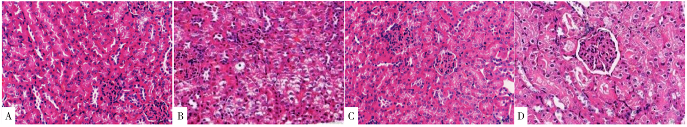
Figure 1 HE staining results of renal tissue in each group
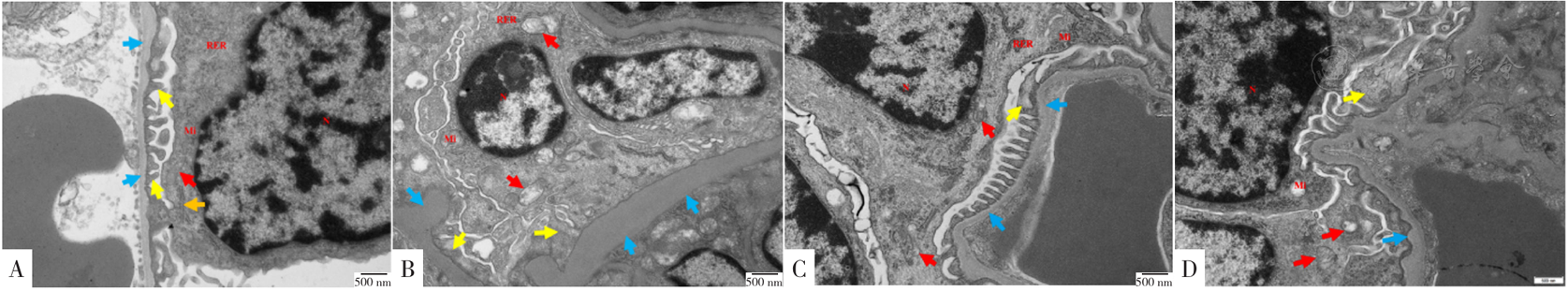
Figure 2 The results of transmission electron microscopy of renal tissue in each group
| 组别 | 只数 | IL-1β | IL-18 |
|---|---|---|---|
| 对照组 | 8 | 10.18±1.90 | 20.85±2.70 |
| 模型组 | 8 | 40.57±4.01a | 87.79±4.24a |
| 红景天苷组 | 8 | 13.97±2.41b | 32.52±3.67b |
| 红景天苷+NLRP3激活剂组 | 8 | 33.08±5.19c | 73.89±7.30c |
| F值 | 141.90 | 497.18 | |
| P值 | <0.001 | <0.001 |
Table 3 Results of serum IL-1β and IL-18 in each group
| 组别 | 只数 | IL-1β | IL-18 |
|---|---|---|---|
| 对照组 | 8 | 10.18±1.90 | 20.85±2.70 |
| 模型组 | 8 | 40.57±4.01a | 87.79±4.24a |
| 红景天苷组 | 8 | 13.97±2.41b | 32.52±3.67b |
| 红景天苷+NLRP3激活剂组 | 8 | 33.08±5.19c | 73.89±7.30c |
| F值 | 141.90 | 497.18 | |
| P值 | <0.001 | <0.001 |
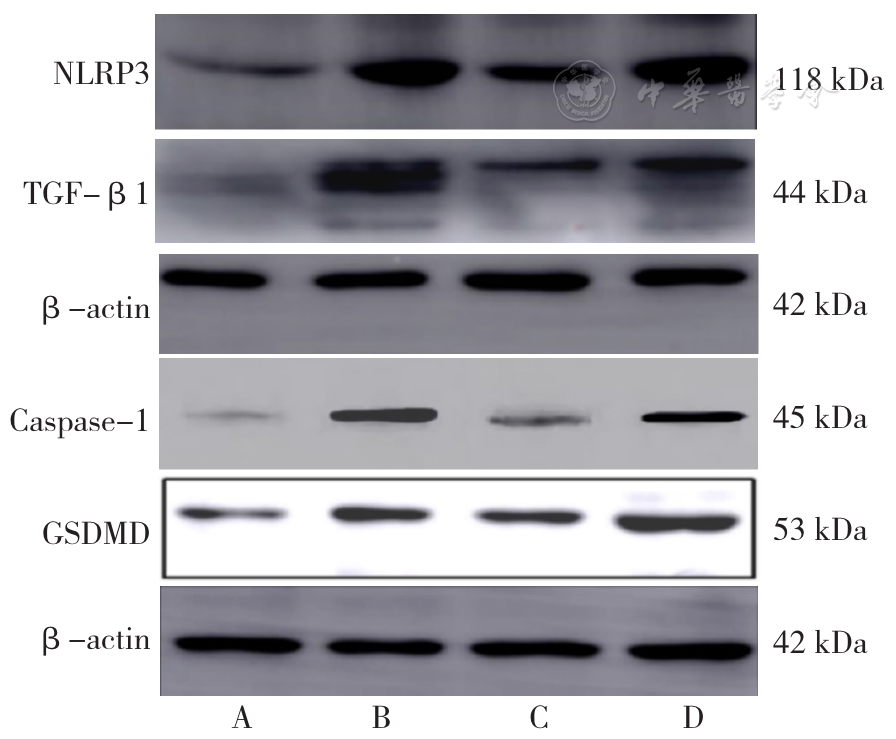
Figure 3 Western bloting protein banding in each group
| 分组 | 只数 | NLRP3 | TGF-β1 | Caspase-1 | GSDMD |
|---|---|---|---|---|---|
| 对照组 | 8 | 0.40±0.06 | 0.21±0.07 | 0.11±0.04 | 0.17±0.03 |
| 模型组 | 8 | 0.97±0.13a | 0.71±0.11a | 0.73±0.08a | 0.52±0.10a |
| 红景天苷组 | 8 | 0.64±0.08b | 0.40±0.09b | 0.18±0.05b | 0.24±0.07b |
| 红景天苷+NLRP3激活剂组 | 8 | 1.04±0.15c | 0.76±0.17c | 0.56±0.07c | 0.56±0.09c |
| F值 | 58.72 | 57.37 | 172.14 | 65.81 | |
| P值 | <0.001 | <0.001 | <0.001 | <0.001 |
Table 4 Protein expression of GSDMD,Caspase-1,NLRP3,and TGF-β1 in each group
| 分组 | 只数 | NLRP3 | TGF-β1 | Caspase-1 | GSDMD |
|---|---|---|---|---|---|
| 对照组 | 8 | 0.40±0.06 | 0.21±0.07 | 0.11±0.04 | 0.17±0.03 |
| 模型组 | 8 | 0.97±0.13a | 0.71±0.11a | 0.73±0.08a | 0.52±0.10a |
| 红景天苷组 | 8 | 0.64±0.08b | 0.40±0.09b | 0.18±0.05b | 0.24±0.07b |
| 红景天苷+NLRP3激活剂组 | 8 | 1.04±0.15c | 0.76±0.17c | 0.56±0.07c | 0.56±0.09c |
| F值 | 58.72 | 57.37 | 172.14 | 65.81 | |
| P值 | <0.001 | <0.001 | <0.001 | <0.001 |
| [1] |
|
| [2] |
|
| [3] |
|
| [4] |
|
| [5] |
|
| [6] |
|
| [7] |
|
| [8] |
|
| [9] | |
| [10] |
|
| [11] |
|
| [12] |
朱四民,王会芳,林凤平,等. 葛根提取物通过调控NOD样受体蛋白3/半胱氨酸天冬氨酸蛋白酶1通路减轻糖尿病大鼠肾损伤的研究[J]. 中国糖尿病杂志,2019,27(11):852-857. DOI:10.3969/j.issn.1006-6187.2019.11.010.
|
| [13] |
|
| [14] |
|
| [15] |
|
| [16] |
|
| [17] |
|
| [18] |
|
| [19] |
|
| [20] |
|
| [21] |
胡龙江,周音频,曹运兰,等. NLRP3-ASC-Caspase-1-IL-18/IL-1β-TGF-β信号通路与Ⅱ型糖尿病临床相关性研究[J]. 西部医学,2018,30(3):335-340,346. DOI:10.3969/j.issn.1672-3511.2018.03.005.
|
| [22] |
|
| [23] |
|
| [24] |
朴敏虎,王程瑜,李香丹,等. 红景天苷对糖尿病肾病大鼠的治疗作用及其机制探讨[J]. 山东医药,2017,57(10):34-36. DOI:10.3969/j.issn.1002-266X.2017.10.010.
|
| Viewed | ||||||
|
Full text |
|
|||||
|
Abstract |
|
|||||