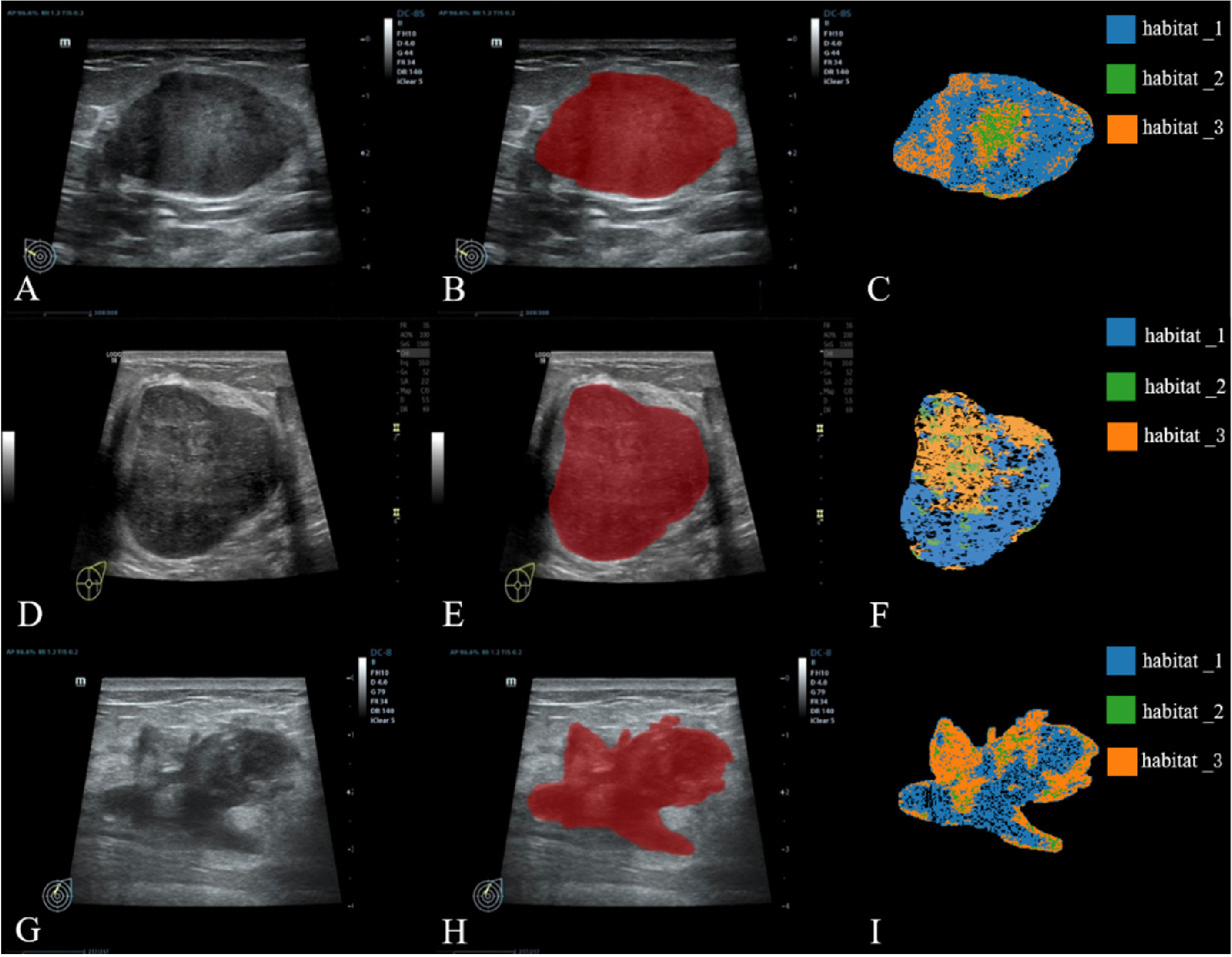
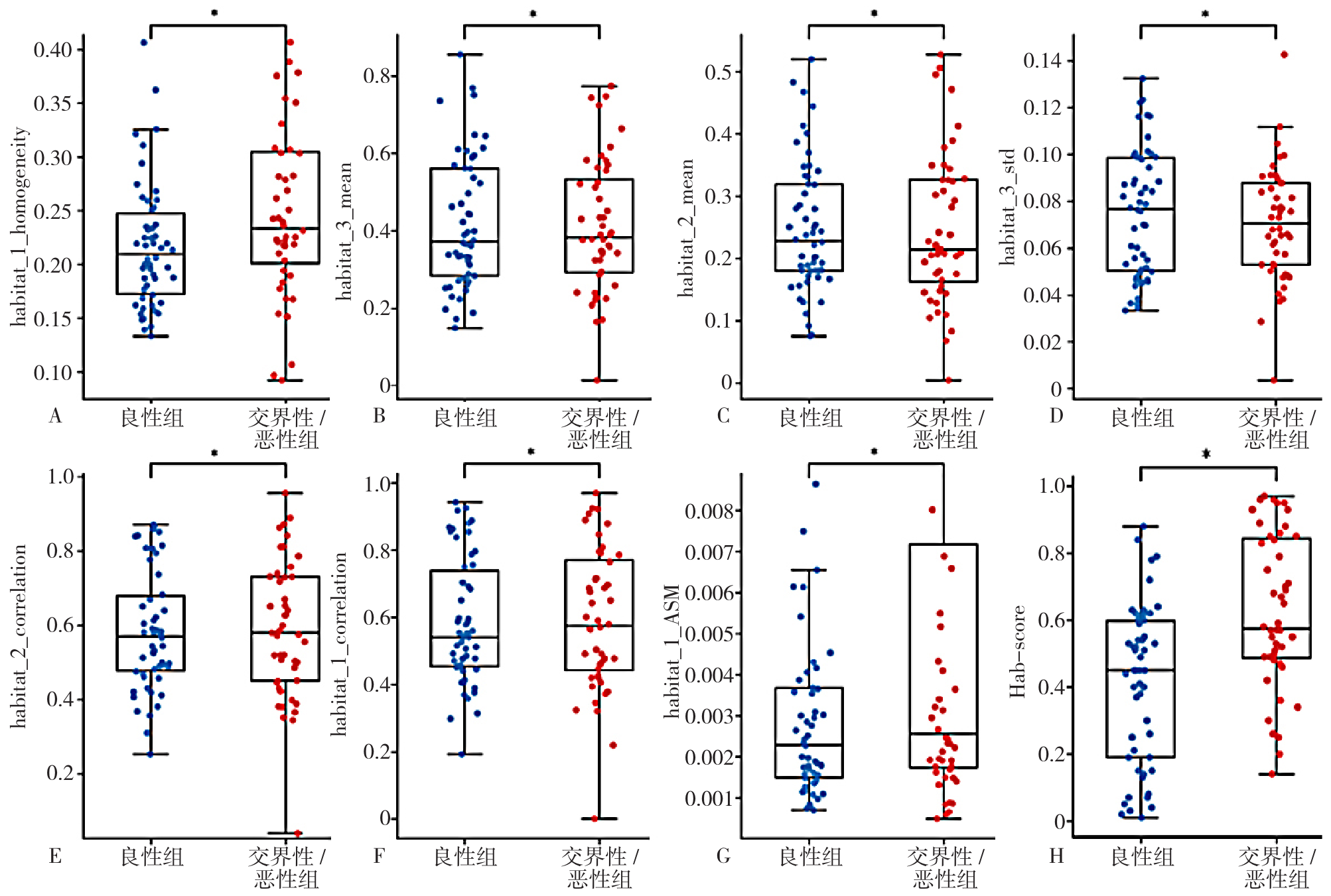
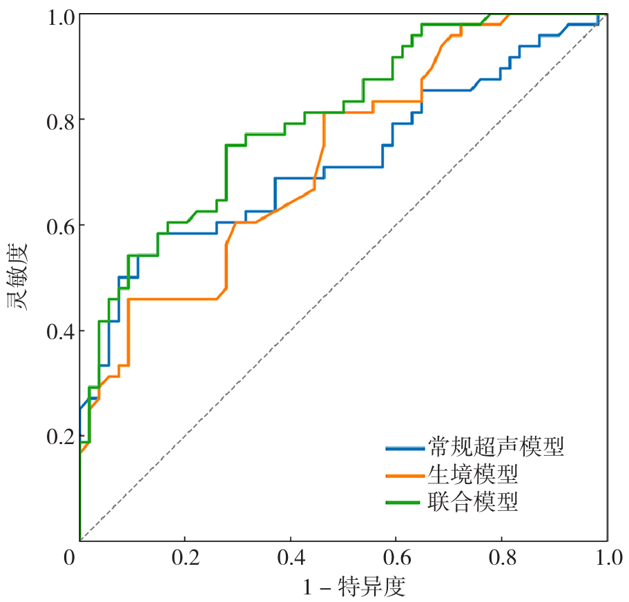
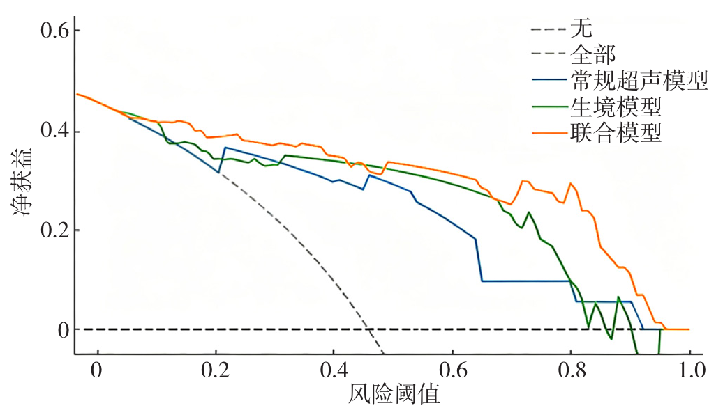
| [1] |
TAN P H, ELLIS I, ALLISON K, et al. The 2019 World Health Organization classification of tumours of the breast[J]. Histopathology, 2020, 77(2): 181-185. DOI: 10.1111/his.14091.
|
| [2] |
陈畅, 孙强, 李炎. 中国女性乳腺分叶状肿瘤诊治专家共识[J]. 中国研究型医院, 2023, 10(4): 1-14.
|
| [3] |
NIU S H, HUANG J H, LI J, et al. Differential diagnosis between small breast Phyllodes tumors and fibroadenomas using artificial intelligence and ultrasound data[J]. Quant Imaging Med Surg, 2021, 11(5): 2052-2061. DOI: 10.21037/qims-20-919.
|
| [4] |
|
| [5] |
LIU Z N, MOUNI D, ZHANG S M, et al. Predicting the early response to neoadjuvant chemotherapy in high-grade serous ovarian cancer by intratumoral habitat heterogeneity based on 18F-FDG PET/CT[J]. Eur J Nucl Med Mol Imaging, 2026, 53(2): 979-991. DOI: 10.1007/s00259-025-07480-z.
|
| [6] |
GONG Z J, LIU Z X, HUANG K Y, et al. Habitat analysis based on magnetic resonance imaging for the prediction of prostate cancer: a dual-center study[J]. Quant Imaging Med Surg, 2025, 15(9): 8395-8408. DOI: 10.21037/qims-2025-223.
|
| [7] |
WANG Y, DAI A, WEN Y, et al. Prediction of high-risk capsule characteristics for recurrence of pleomorphic adenoma in the parotid gland based on habitat imaging and peritumoral radiomics: a two-center study[J]. Acad Radiol, 2025, 32(7): 4134-4145. DOI: 10.1016/j.acra.2025.01.030.
|
| [8] |
ADLER D D, CARSON P L, RUBIN J M, et al. Doppler ultrasound color flow imaging in the study of breast cancer: preliminary findings[J]. Ultrasound Med Biol, 1990, 16(6): 553-559. DOI: 10.1016/0301-5629(90)90020-d.
|
| [9] |
|
| [10] |
PACKER M D C, LESTER S C. Current understanding of phyllodes tumors of the breast: tumor classification, molecular landscape, and best pathology practice[J]. Hum Pathol, 2025, 162: 105863. DOI: 10.1016/j.humpath.2025.105863.
|
| [11] |
BASARA AKIN I, OZGUL H, SIMSEK K, et al. Texture analysis of ultrasound images to differentiate simple fibroadenomas from complex fibroadenomas and benign phyllodes tumors[J]. J Ultrasound Med, 2020, 39(10): 1993-2003. DOI: 10.1002/jum.15304.
|
| [12] |
曹艺敏, 周少萍, 胡田, 等. 乳腺叶状肿瘤良恶性的超声图像特点分析[J]. 医学综述, 2018, 24(24): 4970-4973.
|
| [13] |
LI T, LI Y, YANG Y, et al. Logistic regression analysis of ultrasound findings in predicting the malignant and benign phyllodes tumor of breast[J]. PLoS One, 2022, 17(3): e0265952. DOI: 10.1371/journal.pone.0265952.
|
| [14] |
TSUCHIYA M, MASUI T, TERAUCHI K, et al. MRI-based radiomics analysis for differentiating phyllodes tumors of the breast from fibroadenomas[J]. Eur Radiol, 2022, 32(6): 4090-4100. DOI: 10.1007/s00330-021-08510-8.
|
| [15] |
JING S, WANG H, LIN P, et al. Quantifying and interpreting biologically meaningful spatial signatures within tumor microenvironments[J]. NPJ Precis Oncol, 2025, 9(1): 68. DOI: 10.1038/s41698-025-00857-1.
|
| [16] |
ZHANG X, CHEN X, FU Y, et al. Study on heterogeneity of vascularity and cellularity via multiparametric MRI habitat imaging in breast cancer[J]. BMC Med Imaging, 2025, 25(1):159. DOI: 10.1186/s12880-025-01698-x.
|
| [17] |
XU R, YU D, LUO P, et al. Do habitat MRI and fractal analysis help distinguish triple-negative breast cancer from non-triple-negative breast carcinoma[J]. Can Assoc Radiol J, 2024, 75(3): 584-592. DOI: 10.1177/08465371241231573.
|
| [18] |
|